
Figure 1. High pulse pressure impairs endothelial function in cerebral arteries from young mice, but not old mice.
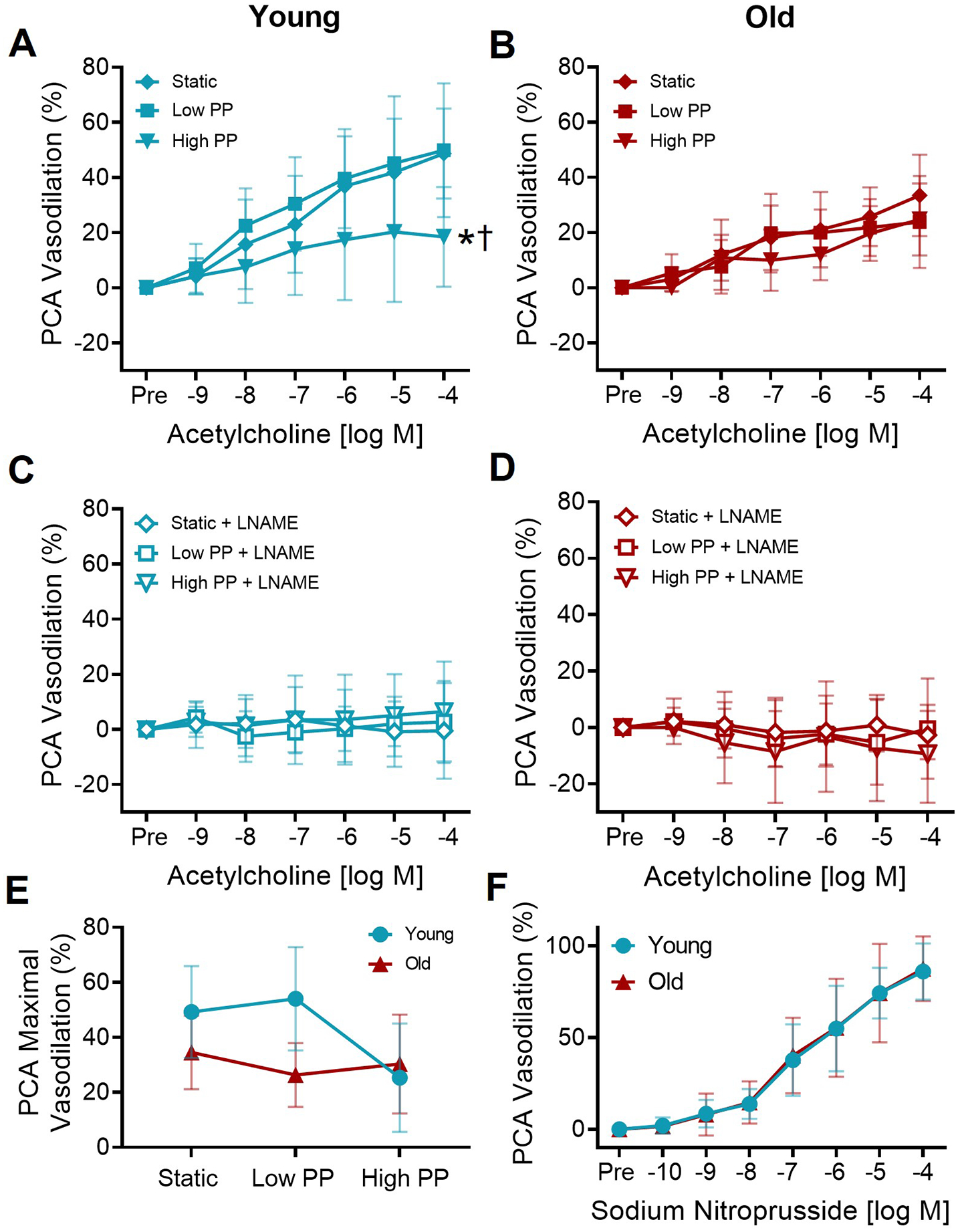
The dose-dependent dilation in (A) young and (B) old posterior cerebral arteries (PCAs) to increasing concentrations of acetylcholine following exposure to static pressure, low pulse pressure (PP), and high PP. In young PCAs, there was a significant ACh dose × PP interaction (p=0.02). There was no ACh dose × PP interaction in the old PCAs. n=6–20/group *p<0.05 high PP vs. static, †p<0.05 high PP vs. low PP, Tukey post-hoc test following significant dose × pulse interaction with RM-ANOVA.. In the presence of nitric oxide synthase inhibitor L-NAME, the dose-dependent dilation in (C) young and (D) old PCAs to increasing concentrations of acetylcholine. n=5–8/group, no interaction of dose × pulse by RM-ANOVA. (C). Maximal dilation to acetylcholine in young and old PCAs following static pressure, low PP, and high PP. n=6–20/group. (D). The dose-dependent dilation of young and old PCAs to increasing concentrations of sodium nitroprusside in PCAs. n=10–11/group, no interaction of dose × age by RM-ANOVA. Values are mean ± SD.