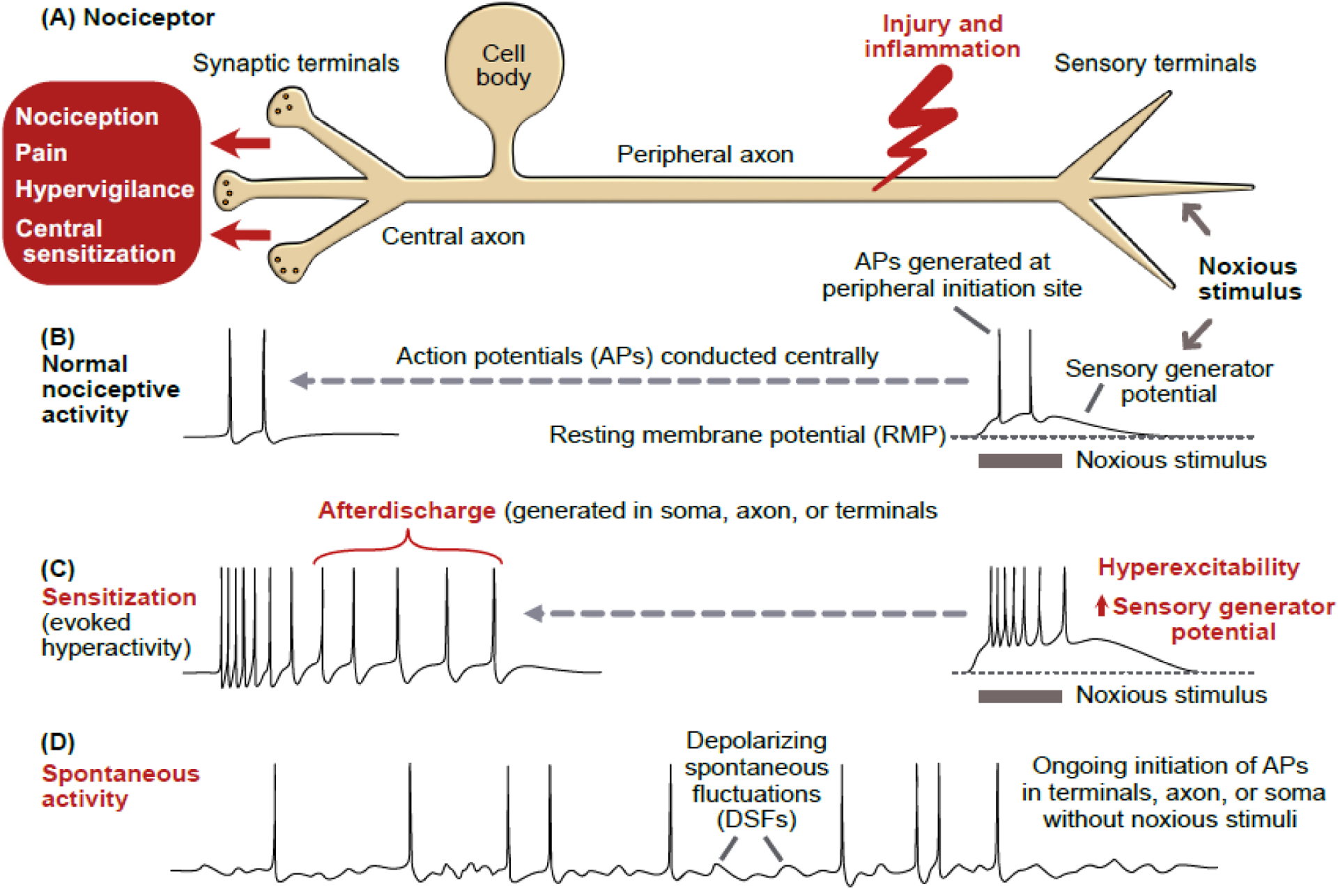
Figure 1. Nociceptor hyperactivity.

(A) Schematic of a primary nociceptor in vertebrates, gastropod molluscs, and leeches. The cell body is centrally located and distant from peripheral terminals where survivable injury and inflammation are most likely. In vertebrates, the cell body is in a ganglion near the central nervous system (CNS). In molluscs and leeches, nociceptor cell bodies are in ganglia within the CNS. (B) Normal nociceptive activity initiated by noxious stimulation of peripheral terminals produces a depolarizing sensory generator potential that may reach threshold for action potentials (APs). These APs are conducted to the CNS, sometimes evoking pain. (C) Illustration of sensitization of nociceptive activity evoked by the same noxious stimulus as in panel B, with the evoked hyperactivity following injury caused by a larger sensory generator potential, increased terminal excitability, and afterdischarge triggered by the evoked APs, which sometimes evoke pain, hypervigilance, and central sensitization. Experimentally, injection of pulses or ramps of depolarizing current (usually into the cell body) is often used to reveal hyperexcitability (see Box 1). (D) Spontaneous activity after injury, defined as ongoing activity generated in the absence of concurrent extrinsic stimulation of a neuron (the spontaneous activity is sometimes generated by depolarizing spontaneous fluctuations, DSFs) [100]. The term spontaneous activity is often used more loosely in referring to any nociceptor activity in the absence of evident ongoing noxious stimulation, including cases where intrinsic (cell autonomous) hyperexcitability and unobserved inflammatory signals combine to drive ongoing hyperactivity. Spontaneous activity and/or afterdischarge have been found in peripheral terminals, injured axons, and cell bodies of mammals and molluscs.
