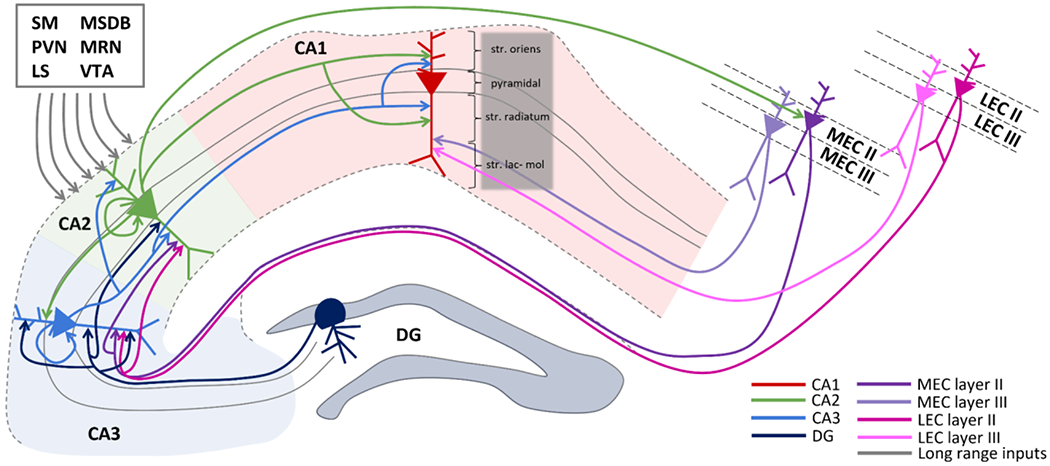
Figure 1: Hippocampal anatomical connectivity.

The main inputs and outputs of area CA2 are shown. CA2 receives projections from CA3, DG, layer II in EC (both medial and lateral portions) and send projections to CA1 and back to layer II in MEC. In addition to intrahippocampal and entorhinal cortex connections, long range inputs from supramammillary nucleus (SM), paraventricular nucleus (PVN), lateral septum (LS), medial septum diagonal band (MSDB), median raphe nucleus (MRN), and ventral tegmental area (VTA) are also included.
