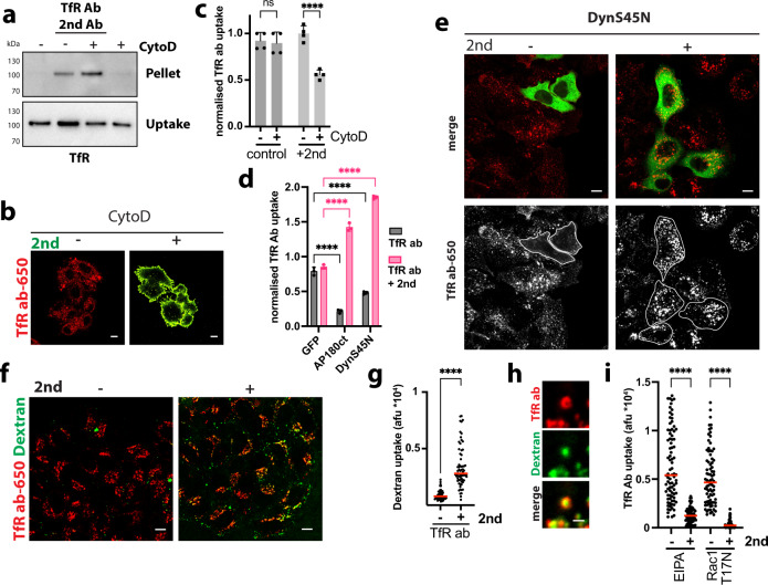
Fig. 4. Aggregation-dependent endocytosis re-routes transferrin receptor.
a Antibody-mediated aggregation and endocytosis of TfR. After surface biotinylation, SkBr3 cells ± CytochalasinD (CytoD) were incubated with anti-transferrin-receptor antibody and cross-linking secondary (2nd) antibody for 30 min, surface remaining biotin was removed. Samples were processed, as described in the “Methods” section and assayed by immunoblot for transferrin receptor (TfR). b, c Cross-linked transferrin-receptor endocytosis requires actin polymerisation. SkBr3 cells ± CytoD were incubated with dylight650-labelled anti-transferrin-receptor antibody 289 (red) ± secondary antibody-AlexaFluor488 (green) (2nd) for 30 min and analysed by confocal microscopy (b) or flow cytometry (c) means ± SD, n = 4 independent experiments, ns (non-significant) P > 0.05, ****P < 0.0001, two-way ANOVA with Sidak’s multiple comparison test. d, e Endocytosis of cross-linked transferrin receptor is independent of clathrin and dynamin. SkBr3 cells transfected with control (GFP) and dominant-negative AP180ct and DynaminS45N N-terminally GFP-tagged expression constructs were incubated with dylight650-labelled anti-transferrin-receptor antibody ± secondary antibody (2nd) for 30 min and analysed by flow cytometry (d) or confocal microscopy (e). Means ± SD, n = 3 independent experiments, ****P < 0.0001; two-way ANOVA with Dunnett’s multiple comparison test shown in (d). DynS45N transfected cells are outlined in lower panels (e). f, g Cross-linked transferrin-receptor endocytosis stimulates fluid-phase uptake. SkBr3 cells were co-incubated with dextran (70 kDa)-TMR (green) and dylight650-labelled anti-transferrin-receptor antibody (red) ± secondary antibody (2nd) for 30 min and analysed by confocal microscopy. Results are quantified in (g) dots represent measurements from individual cells, red lines indicate the median; n ≥ 50 cells from three independent experiments, ****P < 0.0001, two-tailed unpaired Student’s t test. h Cross-linked transferrin-receptor-positive endocytic carriers exhibit dextran-filled lumina. Cross-linked transferrin-receptor endocytosis is inhibited by 5‐(N‐ethyl‐N‐isopropyl)amiloride (EIPA) and dominant-negative Rac1 (i). HeLa cells transfected with dominant-negative Rac1 (T17N) for 16 h, or treated with 50 µM EIPA for 30 min, were incubated with dylight650-labelled anti-transferrin-receptor antibody ± secondary antibody (2nd) for 30 min. After fixation, surface-bound antibody was counterstained and samples analysed by confocal microscopy. Quantification of anti-transferrin-receptor antibody ± secondary antibody (2nd) endocytosis (after surface subtraction) is shown (dots represent measurements from individual cells, red lines indicate the median; n ≥ 50 from three independent experiments, ****P < 0.0001, one-way ANOVA with Sidak’s multiple comparison test). Scale bars: 10 µm (b, e, f), 1 µm (h). Source data are provided as a Source Data file.

