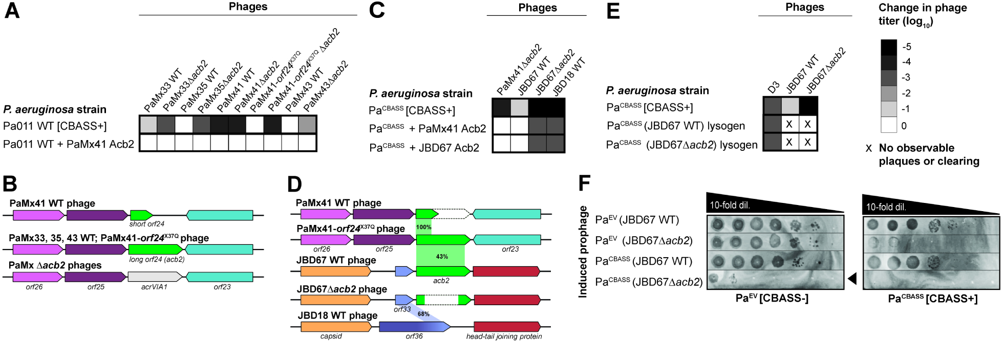
Figure 2. Phage-encoded acb2 is necessary for replication in the presence of CBASS.

(A, C, E) Heat maps representing the order of magnitude change in phage titer, where phage titer is quantified by comparing the number of spots (with plaques, or clearing if plaques were not visible) on the CBASS+ strain divided by the CBASS- strain. Plaque assays used for these quantifications can been seen in Figure S2 (n=3). (B, D) Comparison of the acb2 locus across phage genomes. PaMx41-like Δacb2 phages have the acb2 gene substituted with the type VI-A anti-CRISPR gene (acrVIA1) as part of the knockout procedure, and JBD67Δacb2 phages have the acb2 gene removed from its genome. Genes with known protein functions are indicated with names, and genes with hypothetical proteins are indicated with “orf”. Acb2 percent amino acid identity is shown in (D). (F) Plaque assays assessing the titer induced prophages spotted on a lawn of PaEV or PaCBASS. Black arrowhead highlights reduction in JBD67Δacb2 phage titer. See also Figure S2.
