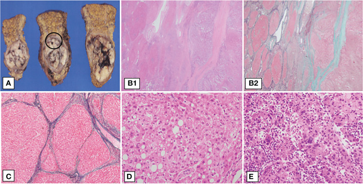
Figure 3.
Pathological findings of the patient’s liver resection specimen. Surgical specimens and the pathological findings. (A) Cut specimen, (B1–D) show magnified images of the black circles. (B1, B2) Liver histological findings of hematoxylin-eosin (H.E) staining and Elastica-Masson (E.M) staining, ×40, respectively. Liver fibrosis was observed, indicating F4 cirrhosis. The left half of the specimen is the cirrhotic area and the right half is the hepatocellular carcinoma area. Hepatocellular carcinoma growing in a macrotrabecular pattern or compact pattern. (C) Liver histological findings of E.M staining, ×100. Hepatocytes show partial fatty degeneration, and the hepatic lobular structure has disappeared, revealing a fibrous septum. (D) Background liver showing fatty degeneration and ballooning of hepatocytes (H&E staining, 400×). (E) Histological findings of hepatocellular carcinoma. The main component is poorly differentiated hepatocellular carcinoma. Tumor cells proliferate in a macrotrabecular pattern and show pleomorphism, such as multinucleation and unequal size (H&E staining, 400×).

