

Abstract
Uterine artery embolization (UAE) is an established technique to treat benign diseases of the uterus such as uterine leiomyomata (fibroids) and adenomyosis. This article reviews the use of UAE in these conditions and summarizes the evidence regarding safety and efficacy of the technique based on the current literature.
Introduction
In recent years, the evidence for uterine artery embolization (UAE) as a therapeutic option for females who seek a uterus preserving alternative to the surgical treatment of uterine leiomyomata and adenomyosis has solidified. While UAE is offered by interventional radiologists in several countries it is still underutilized and unrecognized.1,2 Uterine fibroids and adenomyosis are structural pathologies of the uterus may cause overlapping symptomatology such as hypermenorrhea and dysmenorrhea and can also occur concomitantly.
Current treatment options for uterine leiomyomata
Uterine leiomyomata (also known as uterine fibroids) are the most common type of benign uterine tumors.3–5 Their prevalence increases with age until menopause and this condition affects more females than hypertension or breast cancer.6 There are marked differences by race with higher age-specific incidence and prevalence of uterine fibroids in black females at all ages. Results of observational studies in the UK show that uterine fibroids are under-recorded in primary care.7 The frequency of the condition is likely to be underestimated due to the fact that many females with uterine fibroids are asymptomatic, or symptoms develop insidiously. It has been estimated that uterine fibroids are clinically apparent in 25% of females of reproductive age and cause symptoms severe enough in approximately 25% to require treatment.5 Fibroids can cause heavy menstrual bleeding, pressure symptoms (bulk symptoms), dysmenorrhea and pelvic pain and can impair fertility.8 Fibroids appear to have a greater impact on quality of life than other chronic conditions, including asthma, irritable bowel syndrome, and gastroesophageal reflux disease.9 Many females delay seeking treatment for their fibroids, often for several years, despite the range of non-surgical and minimally invasive treatment options now available. Current invasive management strategies include surgical and non-surgical interventions. Surgical interventions, specifically hysterectomy and uterus preserving techniques such as myomectomy or hysteroscopic resection are the most frequently used to treat symptomatic uterine leiomyomata. Non-surgical approaches include UAE and other image-guided interventions such as high-intensity focused ultrasound (HIFU) among others. Medical treatment options for uterine leiomyomas include agents that address only bleeding symptoms (gonadotropin releasing hormone [GnRH] antagonists, levonorgestrel releasing intrauterine devices [LNG-IUDs], contraceptive steroids, and tranexamic acid) and medications that reduce both bleeding and leiomyoma size (GnRH agonists and selective progesterone receptor modulators). Some of these medical therapies are indicated for long-term use, whereas others are a bridge to surgical treatments, interventional procedures, or menopause.
Uterine artery embolization (UAE) for uterine leiomyomata
First reported in 1995 by Ravina et al, UAE has been proven in several randomized trials to be effective and safe to treat symptomatic uterine fibroids.10–13 Technically, UAE involves catheterization of both uterine arteries and free-flow embolization by particulate material until a desired angiographic endpoint is reached thereby occluding the blood supply to fibroids within the uterus. Rarely, flow is sustained to uterine leiomyomata by enlarged ovarian arteries or other collaterals and need to be addressed by embolization as well14,15 (Figure 1). Spherical and non-spherical permanent embolic particles in the size range of 500–900 micrometer have been tested extensively and used in clinical practice.16 Recently, renewed interest in resorbable embolic agents have led to studies employing these agents in UAE for fibroids.17–20 While UAE for fibroids is per se not a highly selective embolization, the known preferential blood flow to the fibroid plexus vessels downstream to the uterine artery as well as the susceptibility of uterine fibroids to ischemia are factors that lead to (selective) infarction of fibroids while the viability of the myometrium and endometrium is preserved21 (Figure 2). Long-term outcomes are related to the degree of fibroid infarction noted at postembolization MRI, with incomplete fibroid infarction predicting higher short- and midterm recurrence rates.22,23Transient uterine ischemia occurs during UAE as has been demonstrated by serial MR imaging and leads to pain and other side-effects necessitating an individually adaptable pain management protocol.24,25 The procedure is usually performed as an inpatient intervention due to better pain management options and reimbursement issues in many countries but outpatient treatment protocols have been successfully established, including same-day discharge after transradial access.26 No differences in the outcomes of UAE for leiomyomata was observed when performed during different phases of the menstrual cycle.27 A recent systematic review evaluating pain management protocols during UAE found no superiority of one protocol above another in the published literature.28 With respect to peri-interventional antibiotic therapy, no consensus exists. A recent study found that discontinuation of antibiotics given after UAE did not result in an increased rate of infection.29
Figure 1.
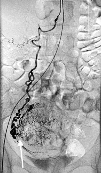
Digital subtraction angiography after selective catherization on the right ovarian artery. The typical downward course of the ovarian artery is depicted with its characteristic corkscrew appearance in its distal segment. The ovarian artery connects to the plexus vessels supplying a uterine leiomyoma (arrow).
Figure 2.

(a-d) T2w and T1w MR images before and after UAE. Multiple well-demarcated hypointense leiomyomata are depicted in (a). T1w contrast-enhanced imaging shows these fibroids are well-perfused except for a hypoperfused area next to the L5/S1 vertebra (white Arrow). A finding that sometimes can be seen in large polyfibroid uteri (B). T2w imaging obtained 6 months after UAE shows that the fibroids have undergone transformation due to infarction and now exhibit a homogenously hypointense signal intensity and moderate reduction in size (C). Contrast-enhanced T1w imaging confirms complete infraction of treated fibroids.
Counseling and interdisciplinary care
In the opinion of the author, females seeking advice for treatment of uterine fibroids should ideally be seen and offered care by an interdisciplinary team of an interventional radiologist and a gynecologist in order to meet the patient’s desire for full disclosure of all treatment options. Multidisciplinarity consultation for patients has been advocated by interventional radiology socities and national expert Groups.30–32 This implies and active role of the interventional radiologist as the care-giver of these patients and resources to schedule outpatient consultation prior and in the follow-up of the intervention. Even in hospitals offering regularly UAE, counseling of patients may be suboptimal if not done in close exchange between interventional radiologists and gynecologists. A study from the Netherlands has shown that misconceptions and information deficits regarding UAE are not uncommon, with nearly half of the gynecologists in hospitals offering UAE overestimating the chances of a surgical intervention after UAE for fibroids.33 Zurawin and Fischer documented nicely, that a “trusting, collaborative, long-term, noncompetitive “win-win” relationship between the gynecologist and radiologist meets the patient’s desire for full disclosure of all myoma treatment options, improves the patient’s overall medical care and physician/patient experience (…)”.34 Patient selection for UAE requires consideration of presenting symptoms, clinical history, size number and location of the leiomyomata or other uterine conditions, patient interest in future fertility, and patient preferences regarding treatment options.30 During the initial clinical visit a physical examination by a gynecologist and a recent pap smear (within a year) should be documented. Approximately 10–30% of fertile females suffer from heavy menstrual bleeding. In 40% of these females uterine fibroids are the cause for this abnormal uterine bleeding (AUB).35 Although relatively uncommon, atypical hyperplasia and malignancy are important potential causes associated with AUB and must be considered in nearly all females of reproductive age. In case of an atypical pattern of uterine bleeding, it is therefore necessary to obtain endometrial tissue sampling. While transvaginal ultrasound is the initial imaging test of choice, MR imaging plays a role to assess the extent of disease, exclude other pathologies and weigh possible advantages or disadvantages of UAE versus other treatment options.36–43
Indications and contraindications
UAE is indicated for the treatment of uterine leiomyomata that are causing significant symptoms such as heavy or prolonged menstrual bleeding (hypermenorrhea), pelvic pressure or pain related to identified leiomyomata, including dyspareunia (Table 1). Most females will experience a combination of symptoms. Interventional radiologists should ensure that these symptoms are related to a history of uterine fibroid disease and in accordance with imaging findings. Only a few absolute contraindications for elective UAE in symptomatic benign uterine leiomyomata exist such as pregnancy, an active and untreated infection or a suspected uterine, cervical or adnexal malignancy. Symptoms and imaging do not allow exclusion of a uterine sarcoma in particular. However, MR imaging can aide in the differentiation of atypical fibroids and uterine sarcomas exist and key features exist, that are more suggestive of a uterine sarcoma.42,43 Coffin et al seeked to determine the frequency of uterine malignancy in patients evaluated for UAE at a single institution. Uterine malignancy was rare (4/864, 0.46%), and MRI detected the majority, 3 (75%) of 4 before UAE.44
Table 1.
Symptoms and complaints associated with uterine fibroids
|
|
|
|
|
UAE is indicated for the treatment of uterine leiomyomata that are causing significant symptoms. Occasionally a single symptom, but more commonly a combination of symptoms
The decision for an organ-preserving medication-based, surgical, or interventional-radiological treatment option should therefore include explanation of the risks of delayed diagnosis of a sarcoma. The spreading of tumor cells as opposed to morcellation after UAE has not been observed. In the case of a lack of response to treatment or a lack of a reduction in the size of the leiomyomata, an insufficient embolization result and the presence of a uterine sarcoma must be considered in the differential diagnosis. Based on a recent A recent retrospective cohort study, who investigated the incidence of gynecologic cancers in adult females who had undergone UAE for fibroids between 2007 and 2017, the authors found that one in 497 women undergoing UAE was diagnosed with a gynecologic malignancy within 3 years after treatment and concluded that short-term malignancies after UAE highlight the importance of pre-procedure evaluation in symptomatic females and females with age-related risk.45
Neither size, number, location, or type of fibroid represent a true contraindication of the procedure.46–49 However, given the wide variations in the extent of fibroid disease, ranging from a single huge leiomyoma, numerous fibroids and those with a distinct anatomical location such as a pedunculated cervical fibroid, every case should be addressed individually. UAE for uterine fibroids should only be carried out after counseling the patient about alternative options, expected benefits of the procedure, possible side effects and complications as well as long-term results of the procedure. This includes but is not limited to a discussion on the individual patient perspective on issues of fertility, uterine preservation, expected shrinkage of fibroids, preference for a certain type of treatment, and risk of subsequent treatments after UAE as well as the risk of missed cancer.30,50,51
Side effects and complications
Large-scale trials and registries have been conducted that underline the safety of UAE for uterine fibroids.52,53 Reported complications include prolonged or poorly controlled pain, infection (pyomyoma, endometritis, or tubo-ovarian abscess), urinary tract infection or urinary retention, and vessel or nerve injury at the access site. Post-embolization syndrome (PES) with pain, fever and nausea as well as elevated inflammatory markers are a common side-effect of solid organ embolization and should not be viewed as a complication of UAE but properly treated. This holds true also for prolonged vaginal discharge and less frequently delayed passage of fibroid material with or without symptoms (Figure 3). Rare major complications include death secondary to sepsis or pulmonary embolism, inadvertent embolization of a leiomyosarcoma, uterine necrosis, buttock necrosis, labial necrosis, vesico-uterine fistula formation, small-bowel volvulus, and acute renal failure. Thresholds for both expected outcomes and complications are given in society guidelines and elsewhere30,31 (Table 2).
Figure 3.

(a,b) T1w and T2w MR images 12 months after UAE. On T1w imaging an intramual/submucosal fibroid with a sharp hypointense, presumably calcified right-sided border is seen. The left sided part shows a broad stripe of hypointense tissue with an irregular hyperintense border(A). On T2w imaging the typical homogenous hypointense signal of the infarcted fibroid is bordered by hyperintense tissue in connection to the uterine cavity (arrow in B). This finding represents an area of sloughing and endometrial overgrowth. The patient reported a non-oderous discharge which was treated conservatively until resumption.
Table 2.
Complications of UAE for Leiomyomata
| Complication | Reported Rate (%) | Suggested Threshold (%) |
|---|---|---|
| Permanent amenorrhea | ||
| Age <45 y | 0–3 | 3 |
| Age >45 y | 20–40 | 45 |
| Prolonged vaginal discharge | 2–17 | 20 |
| Transcervical leiomyoma expulsion | 3–15 | 15 |
| Septicemia | 1–3 | 3 |
| DVT/pulmonary embolus | < 1 | 2 |
| Nontarget embolization | < 1 | < 1 |
DVT, deep vein thrombosis; UAE, uterine artery embolization.
Adapted from Dariushnia etal30
Clinical results of Uterine artery embolization (UAE) for uterine leiomyomata
UAE is technically successful in 95–97% of cases, leads to measurable elimination of fibroid-related abnormal uterine bleeding in >90% of treated females, substantial improvement of subjective bulk symptoms and 80 to 90% satisfaction rate among treated females.31 Thresholds for expected outcomes of the procedure have been published (Table 3.) Multiple RCTs have proven UAE to be a safe, effective alternative to surgical treatment for symptomatic uterine fibroids.12,13,54,55,57,58 The latest RCT, comparing UAE to myomectomy showed that both treatments are effective for improving the quality of life of females with symptomatic uterine fibroids, with an early reported advantage of myomectomy over UAE not being sustained at four years.59 The introduction of the Uterine Fibroid Symptom and Quality-of-Life questionnaire by Spies et al in 2002 was an important step toward the collection of meaningful patient-centric outcome data besides safety and durability, allowing to quantify the burden of uterine fibroids on health-related quality of life (HRQOL) and enabling the comparison of different treatments.9,12,60,61
Table 3.
Expected Outcomes of UAE for Leiomyomata
| Outcome | Reported Rate (%) | Threshold (%) |
|---|---|---|
| Leiomyoma size reduction | 50–60 | 40 |
| Uterine size reduction | 40–50 | 30 |
| Reduction of bulk symptoms | 88–92 | 80 |
| Elimination of abnormal uterine bleeding | > 90 | 85 |
| Successful elimination of symptoms | 75 | 70 |
| Patient satisfaction (would recommend UAE to a friend) | 80–90 | 75 |
| Secondary hysterectomy Secondary surgical procedure |
4% at 12 months (REST) 23% at 24 months (EMMY) 11% at 32 months (REST) 28% at 60 months (EMMY) 24% at 48 months (FEMME) |
|
| Patient satisfaction (would recommend UAE to a friend) | 80–90 | 75 |
There have been many studies on comparative outcome between UAE and other uterine preserving treatments, most of them comparing UAE with myomectomy. In a recent systematic review, Cope and Stewart concluded that UAE had similar quality of life scores, symptom severity scores, sexual function scores, ovarian function, and miscarriage rates following intervention in comparison with myomectomy. There was a lower likelihood of conceiving after UAE and a higher rate of re-intervention after UAE compared with myomectomy. However, the magnitude of this difference in re-intervention rate was very small (4%) and only present in studies with a mean of at least 3 years of follow-up.62 There is currently only limited or inconsistent scientific evidence that could inform which procedure should be preferred over another to treat symptomatic leiomyomas in patients who desire uterine preservation for future pregnancy. High intensity focused ultrasound (HIFU) for symptomatic leiomyomata has been investigated in recent years offering the advantages of being non-invasive and outpatient procedure. There is, however, a paucity of data comparing the efficacy of HIFU to UAE. In the FIRSTT study, the only randomized trial comparing HIFU to UAE, a lower reintervention rate and greater improvement in symptoms was seen after uterine artery embolization.63
Areas of uncertainty – Prospect of improving care
Despite a plethora of studies on UAE for fibroids including level I evidence on its safety and efficacy, several areas of uncertainty but at the same time the prospect of improving care exist.
While it is clear that pregnancy is attainable for females undergoing UAE with many of these pregnancies proceed uneventfully to successful deliveries, the actual fertility rate following UAE remains uncertain.64,65 Further randomized trials should include the assessment of the intention for pregnancy and enroll females who desire future pregnancy. Among studies comparing UAE with myomectomy, there is considerable heterogeneity and variation in patient characteristics, extent of fibroid disease, presenting symptoms and for example surgical approach.62 Further studies should therefore address proper defined (sub)groups that may benefit more from a non-extirpative treatment such as females with a high fibroid burden, anaemia or previous myomectomy as these are considered to have a higher risk of perioperative complications after surgery and may benefit especially from UAE.66,67 Same holds true for large (giant) fibroids, which can be effectively treated by UAE but where the limited available data indicate a relatively higher risk of complications and re-interventions and patients may be better treated by myomectomy.68
Current treatment option for adenomyosis of the uterus
Adenomyosis is a commonly encountered benign uterine disease affecting females of reproductive age and is characterized by the presence of ectopic endometrial glands and stroma surrounded by hyperplastic and hypertrophic smooth muscle of the myometrium.69 The pathogenesis and aetiology of adenomyosis remains unknown. Invagination of the basal endometrium as a result of activation of the tissue injury and repair (TIAR) mechanism and metaplasia of displaced embryonic pluripotent Mullerian remnants or differentiation of adult stem cells are theories discussed in the literature.70 The prevalence of adenomyosis is about 20% in premenopausal females.71 Typical symptoms include often debilitating menorrhagia, chronic pelvic pain, and dysmenorrhea. Adenomyosis is associated with a negative impact on fertility, decreasing the rate of spontaneous pregnancy and increasing the rate of abortion.72
The choice of therapy for adenomyosis should take into account the clinical presentation of females and the desire for future pregnancy given the increasing number of nulliparous, younger patients with this condition. Medical management is still controversial and no drug is specifically labeled for use in case of uterine adenomyosis.73 The use of a Levonorgestrel intrauterine device (LNG-IUD) has been reported to be successful.74 LNG-IUD seem to be most optimal in females with moderate uterine enlargement.75 Until recently, hysterectomy has been the only definitive treatment for females with adenomyosis who have completed child-bearing. Uterine preserving surgical techniques are under investigation but have not been widely adopted outside Japan.76,77 By debulking or excision these techniques aim to remove diseased tissue while preserving the integrity of the uterus. A recent systematic review and meta-analysis on the outcome of fertility-sparing and nonfertility-sparing surgery for the treatment of adenomyosis summarized that uterine preserving surgical treatment of adenomyosis results in high rates of control of symptoms for pain (>70% at 12 months) and bleeding (>70% at 12 months), and in many cases facilitates conception without endangering the outcome of pregnancy.78 However, none of the techniques can guarantee complete excision of the disease from within the myometrium due to its infiltrative pattern and these procedures increase the risk of uterine rupture in case of pregnancy. The associated risk of uterine rupture after surgery which has been reported to be between 2–8% after removal of localized uterine adenomyosis compares to 0.26% in pregnancies following myomectomy.77,79 Ablative techniques such as endometrial ablation are effective for superficial adenomyosis but carry a high rate of recurrence for deep infiltrating adenomyosis of around 25% and subsequent hysterectomy in 19% within 3 years after treatment.80 The results of studies employing MR- or ultrasound-guided high-intensity focused ultrasound (HIFU) to treat focal and diffuse adenomyosis have been analyzed in a recent systematic review and meta-analysis. Within a maximum follow-up period of 40 months these studies demonstrated that HIFU is effective in relieving dysmenorrhea in the range of 84% and improving quality of life as well as reducing uterine volume (reduction rate: 33.6%) and volume of the treated adenomyosis (reduction rate: 45.1%).81 Limitations of the technique include availability, overall cost, unknown fertility outcomes, and anatomical restrictions such as size of the adenomyosis, abdominal scar tissue/pelvic adhesions, body weight and other issues impeding beam transmission.
Uterine artery embolization (UAE) for adenomyosis of the uterus
Early research on UAE for isolated adenomyosis was deemed of questionable clinical benefit with only 55% of treated patients showing sustained clinical improvement after 2 years.82 However, several small and medium-sized case series reported continuously encouraging results with respect to dysmenorrhea and menorrhagia, reduction in the size of the uterus and treated adenomyosis as well as improvement in the quality of life of females who underwent UAE.83–93 Technically, UAE for adenomyosis is similar to UAE for fibroids with the difference that an aggressive endpoint like complete stasis of flow within the uterine arteries is the accepted endpoint. Modifications of the technique have been published using smaller particles with subsequent upsizing.94,95 Pain after UAE for adenomyosis has been referred to be more intense and therfore UAE maybe carried out under epidural anesthesia or patient controlled analgesia (PCA). A systematic review and meta-analysis showed that long-term (> 12 months) improvement of symptoms after UAE for pure (isolated) adenomyosis is 74 and 85.4% for adenomyosis combined with leiomyomata of the uterus.96 In larger case series with up to 7 years follow-up, 82% of UAE treated patients with symptomatic adenomyosis sustained improvement in health-related quality of life as well as control of symptoms was achieved and hysterectomy avoided.97 Comparative studies between UAE and other uterine preserving or medical therapies are lacking. The protocol for a multicenter randomized trial investigating the impact of UAE on Health-Related Quality of Life (HRQOL) in comparison to hysterectomy has been published but results have not been disclosed yet98
Figure 4.

(a, b) T1w MR images before (a) and 12 months after (b) UAE for symptomatic adenomyosis. The uterus is asymmetrically enlarged. The junctional zone of the posterior wall is thickened and hyperintense spots as wells as claw-like protrusion extend from the endometrium into the myometrium (arrow in A). MR imaging 12 months later shows reduction in size of the uterus and the adenomyosis, with a hypointense area corresponding to infarcted adenomyotic tissue is seen in the posterior uterine wall. The patient reported a marked improvement regarding her bleeding symptoms (hypermenorrhea and dysmenorrhea).
Counseling and interdisciplinary care
While many aspects of the counselling for UAE in patients with leiomyoma and adenomyosis overlap, several important differences exist. Adenomyosis was long considered a disease of middle-aged parous females (40–50 years) based on the evaluation of hysterectomy specimen.99 It should be noted, that more recent research show that adenomyosis is a multifaceted disease often diagnosed by non-invasive techniques in younger females with abnormal uterine bleeding (AUB), infertility or pelvic pain.100,101 In 15–57% of the cases, uterine fibroids and adenomyosis coexist in the same uterus and females with both conditions are more likely to experience pelvic pain.102 While uterine fibroids are usually a straightforward diagnosis on ultrasound, adenomyosis especially with concomitant leiomyomata can be challenging and the preoperative diagnosis of adenomyosis remains poor.101 Furthermore, deep infiltrating endometriosis (DIE) is frequently associated with adenomyosis and adds to the difficulty of diagnosis.103,104 In summary, there is interest in therapies that may potentially not only preserve fertility but also avoid major surgery. Thus, careful interdisciplinary evaluation is necessary prior to offering treatment.
Indications, contraindications, side effects and complications
While for UAE for fibroids commonly accepted indications and contraindications exist, there are no evidence-based recommendations for UAE in adenomyosis. Generally, females with symptomatic pure adenomyosis or dominant adenomyosis when both adenomyosis and fibroids coexist and females with an indication for hysterectomy (either failed or refused medical treatment) may be offered the procedure. Exclusion criteria are patients under 18 years of age, pelvic infection, suspected or confirmed malignancy, current or future desire to conceive and concomitant deep infiltrating endometriosis requiring surgery. No specific complications differing from those seen in UAE for leiomyomata have been reported for UAE for adenomyosis.
Areas of uncertainty – Prospect of improving care
UAE for adenomyosis shows favorable clinical outcomes, but randomized controlled trials (RCT) are still lacking. Currently, the RCT “Quality of Life after Embolization vs Hysterectomy in Adenomyosis” (QUESTA) trial is ongoing and results highly awaited. Further studies need to evaluate the impact of UAE on fertility and pregnancy rates in females with adenomyosis and the use of Levonorgestrel intrauterine device (LNG-IUD) to prevent recurrent disease in those, who do not seek pregnancy. The association of adenomyosis with and the impact of endometriosis need further evaluation. MR imaging has established different types of adenomyosis.56 Most of the known imaging features have not been correlated yet with the clinical presentation of adenomyosis, thus their diagnostic and prognostic value is still unknown. A classification system integrating ultrasound and MR imaging findings has not been developed but could help not only to diagnose adenomyosis accurately but also be helpful in deciding the best treatment modality.105 Further research is needed in order to better understand how different phenotypes of adenomyosis respond to UAE.
REFERENCES
- 1. Geary RS, Gurol-Urganci I, Kiran A, Cromwell DA, Bansi-Matharu L, Shakespeare J, et al. Factors associated with receiving surgical treatment for menorrhagia in England and Wales: findings from a cohort study of the National heavy menstrual bleeding audit. BMJ Open 2019; 9(2): e024260. doi: 10.1136/bmjopen-2018-024260 [DOI] [PMC free article] [PubMed] [Google Scholar]
- 2. Clements W, Ang WC, Law M, Goh GS. Treatment of symptomatic fibroid disease using uterine fibroid embolisation: an Australian perspective. Aust N Z J Obstet Gynaecol 2020; 60: 324–29. doi: 10.1111/ajo.13120 [DOI] [PubMed] [Google Scholar]
- 3. Bulun SE. Uterine fibroids. N Engl J Med 2013; 369: 1344–55. doi: 10.1056/NEJMra1209993 [DOI] [PubMed] [Google Scholar]
- 4. Drayer SM, Catherino WH. Prevalence, morbidity, and current medical management of uterine leiomyomas. Int J Gynaecol Obstet 2015; 131: 117–22. doi: 10.1016/j.ijgo.2015.04.051 [DOI] [PubMed] [Google Scholar]
- 5. Stewart EA. Uterine fibroids. Lancet 2001; 357: 293–98. doi: 10.1016/S0140-6736(00)03622-9 [DOI] [PubMed] [Google Scholar]
- 6. Al-Hendy A, Myers ER, Stewart E. Uterine fibroids: burden and unmet medical need. Semin Reprod Med 2017; 35: 473–80. doi: 10.1055/s-0037-1607264 [DOI] [PMC free article] [PubMed] [Google Scholar]
- 7. Martín-Merino E, Wallander M-A, Andersson S, Soriano-Gabarró M, Rodríguez LAG. The reporting and diagnosis of uterine fibroids in the UK: an observational study. BMC Womens Health 2016; 16: 45. doi: 10.1186/s12905-016-0320-8 [DOI] [PMC free article] [PubMed] [Google Scholar]
- 8. Donnez J, Dolmans MM. Uterine fibroid management: from the present to the future. Hum Reprod Update 2016; 22: 665–86. doi: 10.1093/humupd/dmw023 [DOI] [PMC free article] [PubMed] [Google Scholar]
- 9. Downes E, Sikirica V, Gilabert-Estelles J, Bolge SC, Dodd SL, Maroulis C, et al. The burden of uterine fibroids in five European countries. Eur J Obstet Gynecol Reprod Biol 2010; 152: 96–102. doi: 10.1016/j.ejogrb.2010.05.012 [DOI] [PubMed] [Google Scholar]
- 10. de Bruijn AM, Ankum WM, Reekers JA, Birnie E, van der Kooij SM, Volkers NA, et al. Uterine artery embolization vs hysterectomy in the treatment of symptomatic uterine fibroids: 10-year outcomes from the randomized EMMY trial. Am J Obstet Gynecol 2016; 215: 745. doi: 10.1016/j.ajog.2016.06.051 [DOI] [PubMed] [Google Scholar]
- 11. Edwards RD, Moss JG, Lumsden MA, Wu O, Murray LS, Twaddle S, et al. Uterine-artery embolization versus surgery for symptomatic uterine fibroids. N Engl J Med 2007; 356: 360–70. doi: 10.1056/NEJMoa062003 [DOI] [PubMed] [Google Scholar]
- 12. Manyonda I, Belli A-M, Lumsden M-A, Moss J, McKinnon W, Middleton LJ, et al. Uterine-artery embolization or myomectomy for uterine fibroids. N Engl J Med 2020; 383: 440–51. doi: 10.1056/NEJMoa1914735 [DOI] [PubMed] [Google Scholar]
- 13. Pinto I, Chimeno P, Romo A, et al. Uterine fibroids: uterine artery embolization versus abdominal hysterectomy for treatment--a prospective, randomized, and controlled clinical trial. Radiology. Feb 2003;226(2):425-31. doi: 10.1148/radiol.2262011716 [DOI] [PubMed] [Google Scholar]
- 14. Scheurig-Muenkler C, Poellinger A, Wagner M, Hamm B, Kroencke TJ.. Ovarian artery embolization in patients with collateral supply to symptomatic uterine leiomyomata. Cardiovasc Intervent Radiol. Dec 2011;34(6):1199-207. doi: 10.1007/s00270-010-9991-y [DOI] [PubMed] [Google Scholar]
- 15. Chang S, Lee MS, Kim MD, et al. Inferior Mesenteric Artery Collaterals to the Uterus during Uterine Artery Embolization: Prevalence, Risk Factors, and Clinical Outcomes. J Vasc Interv Radiol. Sep 2013;24(9):1353-60. doi: 10.1016/j.jvir.2013.05.049 [DOI] [PubMed] [Google Scholar]
- 16. Das R, Champaneria R, Daniels JP, Belli AM. Comparison of embolic agents used in uterine artery embolisation: a systematic review and meta-analysis. Cardiovasc Intervent Radiol 2014; 37: 1179–90. doi: 10.1007/s00270-013-0790-0 [DOI] [PubMed] [Google Scholar]
- 17. Bengtsson J, Cwikiel W, Sundgren PC, Karlstam E, Gavier-Widén D, Keussen I. The effects of uterine artery embolization with a new degradable microsphere in an experimental study. Acta Radiol 2017; 58: 1334–41. doi: 10.1177/0284185117694510 [DOI] [PubMed] [Google Scholar]
- 18. Ye Y, Ren Y, Zeng H, He J, Zhong Z, Wu X. Characterization of calibrated gelatin sponge particles in a rabbit renal embolization model. Cardiovasc Intervent Radiol 2019; 42: 1183–91. doi: 10.1007/s00270-019-02224-7 [DOI] [PubMed] [Google Scholar]
- 19. Hacking N, Maclean D, Vigneswaran G, Bryant T, Modi S. Uterine fibroid embolization (UFE) with optisphere: a prospective study of a new, spherical, resorbable embolic agent. Cardiovasc Intervent Radiol 2020; 43: 897–903. doi: 10.1007/s00270-020-02460-2 [DOI] [PubMed] [Google Scholar]
- 20. Sato H, Sonomura T, Onishi S, Koike M, Tanaka R, Ueda S, et al. Comparison of uterine necrosis after uterine artery embolization with soluble gelatin sponge particles or tris-acryl gelatin microspheres in swine. Cardiovasc Intervent Radiol 2021; 44: 1780–89. doi: 10.1007/s00270-021-02905-2 [DOI] [PubMed] [Google Scholar]
- 21. Banu NS, Gaze DC, Bruce H, Collinson PO, Belli AM, Manyonda IT. Markers of muscle ischemia, necrosis, and inflammation following uterine artery embolization in the treatment of symptomatic uterine fibroids. Am J Obstet Gynecol 2007; 196: 213. doi: 10.1016/j.ajog.2006.10.888 [DOI] [PubMed] [Google Scholar]
- 22. Pelage JP, Guaou NG, Jha RC, Ascher SM, Spies JB. Uterine fibroid tumors: long-term MR imaging outcome after embolization. Radiology 2004; 230: 803–9. doi: 10.1148/radiol.2303030111 [DOI] [PubMed] [Google Scholar]
- 23. Kroencke TJ, Scheurig C, Poellinger A, Gronewold M, Hamm B. Uterine artery embolization for leiomyomas: percentage of infarction predicts clinical outcome. Radiology 2010; 255: 834–41. doi: 10.1148/radiol.10090977 [DOI] [PubMed] [Google Scholar]
- 24. Scheurig-Muenkler C, Wagner M, Franiel T, Hamm B, Kroencke TJ. Effect of uterine artery embolization on uterine and leiomyoma perfusion: evidence of transient myometrial ischemia on magnetic resonance imaging. J Vasc Interv Radiol 2010; 21: 1347–53. doi: 10.1016/j.jvir.2010.05.008 [DOI] [PubMed] [Google Scholar]
- 25. Han K, Kim SY, Kim HJ, Kwon JH, Kim GM, Lee J, et al. Nonspherical polyvinyl alcohol particles versus tris-acryl microspheres: randomized controlled trial comparing pain after uterine artery embolization for symptomatic fibroids. Radiology 2021; 298: 458–65. doi: 10.1148/radiol.2020201895 [DOI] [PubMed] [Google Scholar]
- 26. Sher A, Garvey A, Kamat S, et al. Single-System Experience With Outpatient Transradial Uterine Artery Embolization: Safety, Feasibility, Outcomes, and Early Rates of Return. AJR Am J Roentgenol. Apr 2021;216(4):975-980. doi: 10.2214/AJR.20.23343 [DOI] [PubMed] [Google Scholar]
- 27. Katsumori T, Yoshikawa T, Sasakura Y, Yasumura T, Hisano M.. Timing of Uterine Artery Embolization for Leiomyoma during the Menstrual Cycle. J Vasc Interv Radiol. Mar 2021;32(3):332-338. doi: 10.1016/j.jvir.2020.11.014 [DOI] [PubMed] [Google Scholar]
- 28. Saibudeen A, Makris GC, Elzein A, et al. Pain Management Protocols During Uterine Fibroid Embolisation: A Systematic Review of the Evidence. Cardiovasc Intervent Radiol. Dec 2019;42(12):1663-1677. doi: 10.1007/s00270-019-02327-1 [DOI] [PubMed] [Google Scholar]
- 29. Graif A, Leung DA, McKenna G, Patel KD, Holmes LE, Grilli CJ.. Evaluation of the Effect of Routine Antibiotic Administration after Uterine Artery Embolization on Infection Rates. J Vasc Interv Radiol. Aug 2020;31(8):1263-1269. doi: 10.1016/j.jvir.2020.03.026 [DOI] [PubMed] [Google Scholar]
- 30. Dariushnia SR, Nikolic B, Stokes LS, Spies JB. Society of interventional radiology standards of practice C. quality improvement guidelines for uterine artery embolization for symptomatic leiomyomata. J Vasc Interv Radiol 2014; 25: 1737–47. doi: 10.1016/j.jvir.2014.08.029 [DOI] [PubMed] [Google Scholar]
- 31. van Overhagen H, Reekers JA. Uterine artery embolization for symptomatic leiomyomata. Cardiovasc Intervent Radiol 2015; 38: 536–42. doi: 10.1007/s00270-014-1031-x [DOI] [PubMed] [Google Scholar]
- 32. Kröncke T, David M. Uterine artery embolization (UAE) for fibroid treatment-results of the 7th radiological gynecological expert meeting. Rofo 2019; 191: 630–34. doi: 10.1055/a-0884-3168 [DOI] [PMC free article] [PubMed] [Google Scholar]
- 33. de Bruijn AM, Huisman J, Hehenkamp WJK, Lohle PNM, Reekers JA, Timmermans A, et al. Implementation of uterine artery embolization for symptomatic fibroids in the Netherlands: an inventory and preference study. CVIR Endovasc 2019; 2: 18. doi: 10.1186/s42155-019-0061-5 [DOI] [PMC free article] [PubMed] [Google Scholar]
- 34. Zurawin RK, Fischer JH, Amir L. The effect of a gynecologist-interventional radiologist relationship on selection of treatment modality for the patient with uterine myoma. J Minim Invasive Gynecol 2010; 17: 214–21. doi: 10.1016/j.jmig.2009.12.015 [DOI] [PubMed] [Google Scholar]
- 35. Liu Z, Doan QV, Blumenthal P, Dubois RW.. A systematic review evaluating health-related quality of life, work impairment, and health-care costs and utilization in abnormal uterine bleeding. Value Health. May-Jun 2007;10(3):183-94. doi: 10.1111/j.1524-4733.2007.00168.x [DOI] [PubMed] [Google Scholar]
- 36. Barral M, Place V, Dautry R, et al. Magnetic resonance imaging features of uterine sarcoma and mimickers. Abdom Radiol (NY). Jun 2017;42(6):1762-1772. doi: 10.1007/s00261-017-1076-9 [DOI] [PubMed] [Google Scholar]
- 37. Dueholm M, Lundorf E, Hansen ES, Ledertoug S, Olesen F.. Accuracy of magnetic resonance imaging and transvaginal ultrasonography in the diagnosis, mapping, and measurement of uterine myomas. Am J Obstet Gynecol. Mar 2002;186(3):409-15. doi: 10.1067/mob.2002.121725 [DOI] [PubMed] [Google Scholar]
- 38. Kirby JM, Burrows D, Haider E, Maizlin Z, Midia M.. Utility of MRI before and after uterine fibroid embolization: why to do it and what to look for. Cardiovasc Intervent Radiol. Aug 2011;34(4):705-16. doi: 10.1007/s00270-010-0029-2 [DOI] [PubMed] [Google Scholar]
- 39. Kubik-Huch RA, Weston M, Nougaret S, et al. European Society of Urogenital Radiology (ESUR) Guidelines: MR Imaging of Leiomyomas. Eur Radiol. Aug 2018;28(8):3125-3137. doi: 10.1007/s00330-017-5157-5 [DOI] [PMC free article] [PubMed] [Google Scholar]
- 40. Rajan DK, Margau R, Kroll RR, et al. Clinical utility of ultrasound versus magnetic resonance imaging for deciding to proceed with uterine artery embolization for presumed symptomatic fibroids. Clin Radiol. Jan 2011;66(1):57-62. doi: 10.1016/j.crad.2010.08.005 [DOI] [PubMed] [Google Scholar]
- 41. Siddiqui N, Nikolaidis P, Hammond N, Miller FH. Uterine artery embolization: pre- and post-procedural evaluation using magnetic resonance imaging. Abdom Imaging 2013; 38: 1161–77. doi: 10.1007/s00261-013-9990-y [DOI] [PubMed] [Google Scholar]
- 42. Smith J, Zawaideh JP, Sahin H, Freeman S, Bolton H, Addley HC. Differentiating uterine sarcoma from leiomyoma: BET (1) T (2) ER check! Br J Radiol 2021; 94: 1125. doi: 10.1259/bjr.20201332 [DOI] [PMC free article] [PubMed] [Google Scholar]
- 43. Aminzadeh P, Alibrahim E, Dobrotwir A, Paul E, Goergen S. Multiparametric Mr evaluation of uterine leiomyosarcoma and stump versus leiomyoma in symptomatic women planned for high frequency focussed ultrasound: accuracy of imaging parameters and interobserver agreement for identification of malignancy. Br J Radiol 2021; 94: 1119: 20200483. doi: 10.1259/bjr.20200483 [DOI] [PMC free article] [PubMed] [Google Scholar]
- 44. Coffin PH, Ascher S, Spies J. The risk of uterine malignancy in a population being evaluated for uterine fibroid embolization. J Comput Assist Tomogr 2020; 44: 893–900. doi: 10.1097/RCT.0000000000001104 [DOI] [PubMed] [Google Scholar]
- 45. Bronico JVR, Matthews BJ, Perkins RB, Lee E-M, Morgan JR, Nitschmann CC, et al. Incidence of gynecologic cancers in women after uterine artery embolization. J Minim Invasive Gynecol 2021; 28: 1231–36: S1553-4650(20)31103-1. doi: 10.1016/j.jmig.2020.10.015 [DOI] [PMC free article] [PubMed] [Google Scholar]
- 46. Kido A, Monma C, Togashi K, et al. Uterine arterial embolization for the treatment of diffuse leiomyomatosis. J Vasc Interv Radiol. May 2003;14(5):643-7. doi: 10.1097/01.rvi.0000071095.76348.e0 [DOI] [PubMed] [Google Scholar]
- 47. Margau R, Simons ME, Rajan DK, et al. Outcomes after uterine artery embolization for pedunculated subserosal leiomyomas. J Vasc Interv Radiol. May 2008;19(5):657-61. doi: 10.1016/j.jvir.2007.11.022 [DOI] [PubMed] [Google Scholar]
- 48. Scheurig C, Islam T, Zimmermann E, Hamm B, Kroencke TJ.. Uterine artery embolization in patients with symptomatic diffuse leiomyomatosis of the uterus. J Vasc Interv Radiol. Feb 2008;19(2 Pt 1):279-84. doi: 10.1016/j.jvir.2007.10.017 [DOI] [PubMed] [Google Scholar]
- 49. Smeets AJ, Nijenhuis RJ, van Rooij WJ, Weimar EAM, Boekkooi PF, Lampmann LEH, et al. Uterine artery embolization in patients with a large fibroid burden: long-term clinical and Mr follow-up. Cardiovasc Intervent Radiol 2010; 33: 943–48. doi: 10.1007/s00270-009-9793-2 [DOI] [PMC free article] [PubMed] [Google Scholar]
- 50. Karlsen K, Hrobjartsson A, Korsholm M, Mogensen O, Humaidan P, Ravn P.. Fertility after uterine artery embolization of fibroids: a systematic review. Arch Gynecol Obstet. Jan 2018;297(1):13-25. doi: 10.1007/s00404-017-4566-7 [DOI] [PubMed] [Google Scholar]
- 51. Sandberg EM, Tummers FHMP, Cohen SL, van den Haak L, Dekkers OM, Jansen FW. Reintervention risk and quality of life outcomes after uterine-sparing interventions for fibroids: a systematic review and meta-analysis. Fertil Steril 2018; 109: 698–707. S0015-0282(17)32103-9. doi: 10.1016/j.fertnstert.2017.11.033 [DOI] [PubMed] [Google Scholar]
- 52. Goodwin SC, Spies JB, Worthington-Kirsch R, Peterson E, Pron G, Li S, et al. Uterine artery embolization for treatment of leiomyomata: long-term outcomes from the fibroid registry. Obstet Gynecol 2008; 111: 22–33. doi: 10.1097/01.AOG.0000296526.71749.c9 [DOI] [PubMed] [Google Scholar]
- 53. Dutton S, Hirst A, McPherson K, Nicholson T, Maresh M. A UK multicentre retrospective cohort study comparing hysterectomy and uterine artery embolisation for the treatment of symptomatic uterine fibroids (hopeful study): main results on medium-term safety and efficacy. BJOG 2007; 114: 1340–51. doi: 10.1111/j.1471-0528.2007.01526.x [DOI] [PubMed] [Google Scholar]
- 54. Hehenkamp WJK, Volkers NA, Birnie E, Reekers JA, Ankum WM. Symptomatic uterine fibroids: treatment with uterine artery embolization or hysterectomy -- results from the randomized clinical embolisation versus hysterectomy (EMMY) trial. Radiology 2008; 246: 823–32. doi: 10.1148/radiol.2463070260 [DOI] [PubMed] [Google Scholar]
- 55. Mara M, Fucikova Z, Maskova J, Kuzel D, Haakova L. Uterine fibroid embolization versus myomectomy in women wishing to preserve fertility: preliminary results of a randomized controlled trial. Eur J Obstet Gynecol Reprod Biol 2006; 126: 226–33. doi: 10.1016/j.ejogrb.2005.10.008 [DOI] [PubMed] [Google Scholar]
- 56. Bazot M, Daraï E. Role of transvaginal sonography and magnetic resonance imaging in the diagnosis of uterine adenomyosis. Fertil Steril 2018; 109: 389–97. doi: 10.1016/j.fertnstert.2018.01.024 [DOI] [PubMed] [Google Scholar]
- 57. Manyonda IT, Bratby M, Horst JS, Banu N, Gorti M, Belli AM.. Uterine artery embolization versus myomectomy: impact on quality of life--results of the FUME (Fibroids of the Uterus: Myomectomy versus Embolization) Trial. Cardiovasc Intervent Radiol. Jun 2012;35(3):530-6. doi: 10.1007/s00270-011-0228-5 [DOI] [PubMed] [Google Scholar]
- 58. Moss JG, Cooper KG, Khaund A, et al. Randomised comparison of uterine artery embolisation (UAE) with surgical treatment in patients with symptomatic uterine fibroids (REST trial): 5-year results. BJOG. Jul 2011;118(8):936-44. doi: 10.1111/j.1471-0528.2011.02952.x [DOI] [PubMed] [Google Scholar]
- 59. Daniels J, Middleton LJ, Cheed V, et al. Uterine artery embolization or myomectomy for women with uterine fibroids: Four-year follow-up of a randomised controlled trial. Eur J Obstet Gynecol Reprod Biol X. Jan 2022;13:100139. doi: 10.1016/j.eurox.2021.100139 [DOI] [PMC free article] [PubMed] [Google Scholar]
- 60. Spies JB, Coyne K, Guaou Guaou N, Boyle D, Skyrnarz-Murphy K, Gonzalves SM.. The UFS-QOL, a new disease-specific symptom and health-related quality of life questionnaire for leiomyomata. Obstet Gynecol. Feb 2002;99(2):290-300. doi: 10.1016/s0029-7844(01)01702-1 [DOI] [PubMed] [Google Scholar]
- 61. Wallace K, Zhang S, Thomas L, et al. Comparative effectiveness of hysterectomy versus myomectomy on one-year health-related quality of life in women with uterine fibroids. Fertil Steril. Mar 2020;113(3):618-626. doi: 10.1016/j.fertnstert.2019.10.028 [DOI] [PubMed] [Google Scholar]
- 62. Cope AG, Young RJ, Stewart EA. Non-extirpative treatments for uterine myomas: measuring success. J Minim Invasive Gynecol 2021; 28: 442–52. doi: 10.1016/j.jmig.2020.08.016 [DOI] [PubMed] [Google Scholar]
- 63. Laughlin-Tommaso S, Barnard EP, AbdElmagied AM, Vaughan LE, Weaver AL, Hesley GK, et al. FIRSTT study: randomized controlled trial of uterine artery embolization vs focused ultrasound surgery. Am J Obstet Gynecol 2019; 220: 174. doi: 10.1016/j.ajog.2018.10.032 [DOI] [PMC free article] [PubMed] [Google Scholar]
- 64. Ludwig PE, Huff TJ, Shanahan MM, Stavas JM. Pregnancy success and outcomes after uterine fibroid embolization: updated review of published literature. Br J Radiol 2020; 93: 1105. doi: 10.1259/bjr.20190551 [DOI] [PMC free article] [PubMed] [Google Scholar]
- 65. Serres-Cousine O, Kuijper FM, Curis E, Atashroo D. Clinical investigation of fertility after uterine artery embolization. Am J Obstet Gynecol 2021; 225: 403. doi: 10.1016/j.ajog.2021.05.033 [DOI] [PubMed] [Google Scholar]
- 66. Jansen LJ, Clark NV, Dmello M, Gu X, Einarsson JI, Cohen SL.. Perioperative Outcomes of Myomectomy for Extreme Myoma Burden: Comparison of Surgical Approaches. J Minim Invasive Gynecol. Sep - Oct 2019;26(6):1095-1103. doi: 10.1016/j.jmig.2018.10.022 [DOI] [PubMed] [Google Scholar]
- 67. Liu X, Tang J, Luo Y, Wang Y, Song L, Wang W. Comparison of high-intensity focused ultrasound ablation and secondary myomectomy for recurrent symptomatic uterine fibroids following myomectomy: a retrospective study. BJOG 2020; 127: 1422–28. doi: 10.1111/1471-0528.16262 [DOI] [PubMed] [Google Scholar]
- 68. Llewellyn O, Patel NR, Mallon D, Quinn SD, Hamady M. Uterine artery embolisation for women with giant versus non-giant uterine fibroids: a systematic review and meta-analysis. Cardiovasc Intervent Radiol 2020; 43: 684–93. doi: 10.1007/s00270-019-02359-7 [DOI] [PubMed] [Google Scholar]
- 69. Bergeron C, Amant F, Ferenczy A. Pathology and physiopathology of adenomyosis. Best Pract Res Clin Obstet Gynaecol 2006; 20: 511–21. doi: 10.1016/j.bpobgyn.2006.01.016 [DOI] [PubMed] [Google Scholar]
- 70. García-Solares J, Donnez J, Donnez O, Dolmans M-M. Pathogenesis of uterine adenomyosis: invagination or metaplasia? Fertil Steril 2018; 109: 371–79. doi: 10.1016/j.fertnstert.2017.12.030 [DOI] [PubMed] [Google Scholar]
- 71. Naftalin J, Hoo W, Pateman K, Mavrelos D, Holland T, Jurkovic D. How common is adenomyosis? A prospective study of prevalence using transvaginal ultrasound in a gynaecology clinic. Hum Reprod 2012; 27: 3432–39. doi: 10.1093/humrep/des332 [DOI] [PubMed] [Google Scholar]
- 72. Horton J, Sterrenburg M, Lane S, Maheshwari A, Li TC, Cheong Y. Reproductive, obstetric, and perinatal outcomes of women with adenomyosis and endometriosis: a systematic review and meta-analysis. Hum Reprod Update 2019; 25: 592–632. doi: 10.1093/humupd/dmz012 [DOI] [PubMed] [Google Scholar]
- 73. Vannuccini S, Luisi S, Tosti C, Sorbi F, Petraglia F.. Role of medical therapy in the management of uterine adenomyosis. Fertil Steril. Mar 2018;109(3):398-405. doi: 10.1016/j.fertnstert.2018.01.013 [DOI] [PubMed] [Google Scholar]
- 74. Fedele L, Bianchi S, Raffaelli R, Portuese A, Dorta M.. Treatment of adenomyosis-associated menorrhagia with a levonorgestrel-releasing intrauterine device. Fertil Steril. Sep 1997;68(3):426-9. doi: 10.1016/s0015-0282(97)00245-8 [DOI] [PubMed] [Google Scholar]
- 75. Lee KH, Kim JK, Lee MA, et al. Relationship between uterine volume and discontinuation of treatment with levonorgestrel-releasing intrauterine devices in patients with adenomyosis. Arch Gynecol Obstet. Sep 2016;294(3):561-6. doi: 10.1007/s00404-016-4105-y [DOI] [PubMed] [Google Scholar]
- 76. Oliveira MAP, Crispi CP, Brollo LC, Crispi CP, De Wilde RL.. Surgery in adenomyosis. Arch Gynecol Obstet. Mar 2018;297(3):581-589. doi: 10.1007/s00404-017-4603-6 [DOI] [PubMed] [Google Scholar]
- 77. Osada H. Uterine adenomyosis and Adenomyoma: the surgical approach. Fertil Steril 2018; 109: 406–17. doi: 10.1016/j.fertnstert.2018.01.032 [DOI] [PubMed] [Google Scholar]
- 78. Mikos T, Lioupis M, Anthoulakis C, Grimbizis GF. The outcome of fertility-sparing and nonfertility-sparing surgery for the treatment of adenomyosis. A systematic review and meta-analysis. J Minim Invasive Gynecol 2020; 27: 309–31. doi: 10.1016/j.jmig.2019.08.004 [DOI] [PubMed] [Google Scholar]
- 79. Sizzi O, Rossetti A, Malzoni M, Minelli L, La Grotta F, Soranna L, et al. Italian multicenter study on complications of laparoscopic myomectomy. J Minim Invasive Gynecol 2007; 14: 453–62. doi: 10.1016/j.jmig.2007.01.013 [DOI] [PubMed] [Google Scholar]
- 80. Philip C-A, Le Mitouard M, Maillet L, de Saint-Hilaire P, Huissoud C, Cortet M, et al. Evaluation of novasure® global endometrial ablation in symptomatic adenomyosis: a longitudinal study with a 36 month follow-up. Eur J Obstet Gynecol Reprod Biol 2018; 227: 46–51. doi: 10.1016/j.ejogrb.2018.04.001 [DOI] [PubMed] [Google Scholar]
- 81. Liu L, Wang T, Lei B. Image-Guided thermal ablation in the management of symptomatic adenomyosis: a systematic review and meta-analysis. Int J Hyperthermia 2021; 38: 948–62. doi: 10.1080/02656736.2021.1939443 [DOI] [PubMed] [Google Scholar]
- 82. Pelage J-P, Jacob D, Fazel A, Namur J, Laurent A, Rymer R, et al. Midterm results of uterine artery embolization for symptomatic adenomyosis: initial experience. Radiology 2005; 234: 948–53. doi: 10.1148/radiol.2343031697 [DOI] [PubMed] [Google Scholar]
- 83. Siskin GP, Tublin ME, Stainken BF, Dowling K, Dolen EG. Uterine artery embolization for the treatment of adenomyosis: clinical response and evaluation with MR imaging. AJR Am J Roentgenol 2001; 177: 297–302. doi: 10.2214/ajr.177.2.1770297 [DOI] [PubMed] [Google Scholar]
- 84. Kim MD, Won JW, Lee DY, Ahn CS. Uterine artery embolization for adenomyosis without fibroids. Clin Radiol 2004; 59: 520–26. doi: 10.1016/j.crad.2003.11.018 [DOI] [PubMed] [Google Scholar]
- 85. Kitamura Y, Allison SJ, Jha RC, Spies JB, Flick PA, Ascher SM. Mri of adenomyosis: changes with uterine artery embolization. AJR Am J Roentgenol 2006; 186: 855–64. doi: 10.2214/AJR.04.1661 [DOI] [PubMed] [Google Scholar]
- 86. Kim MD, Kim S, Kim NK, Lee MH, Ahn EH, Kim HJ, et al. Long-Term results of uterine artery embolization for symptomatic adenomyosis. AJR Am J Roentgenol 2007; 188: 176–81. doi: 10.2214/AJR.05.1613 [DOI] [PubMed] [Google Scholar]
- 87. Lohle PNM, De Vries J, Klazen CAH, Boekkooi PF, Vervest HAM, Smeets AJ, et al. Uterine artery embolization for symptomatic adenomyosis with or without uterine leiomyomas with the use of calibrated tris-acryl gelatin microspheres: midterm clinical and MR imaging follow-up. J Vasc Interv Radiol 2007; 18: 835–41. doi: 10.1016/j.jvir.2007.04.024 [DOI] [PubMed] [Google Scholar]
- 88. Bratby MJ, Walker WJ. Uterine artery embolisation for symptomatic adenomyosis -- mid-term results. Eur J Radiol 2009; 70: 128–32. doi: 10.1016/j.ejrad.2007.12.009 [DOI] [PubMed] [Google Scholar]
- 89. Froeling V, Scheurig-Muenkler C, Hamm B, Kroencke TJ. Uterine artery embolization to treat uterine adenomyosis with or without uterine leiomyomata: results of symptom control and health-related quality of life 40 months after treatment. Cardiovasc Intervent Radiol 2012; 35: 523–29. doi: 10.1007/s00270-011-0254-3 [DOI] [PubMed] [Google Scholar]
- 90. Smeets AJ, Nijenhuis RJ, Boekkooi PF, Vervest HAM, van Rooij WJ, Lohle PNM. Long-Term follow-up of uterine artery embolization for symptomatic adenomyosis. Cardiovasc Intervent Radiol 2012; 35: 815–19. doi: 10.1007/s00270-011-0203-1 [DOI] [PubMed] [Google Scholar]
- 91. Wang S, Meng X, Dong Y. The evaluation of uterine artery embolization as a nonsurgical treatment option for adenomyosis. Int J Gynaecol Obstet 2016; 133: 202–5. doi: 10.1016/j.ijgo.2015.09.016 [DOI] [PubMed] [Google Scholar]
- 92. Yuan K, Zhang JL, Yan JY, et al. Uterine Artery Embolization with Small-Sized Particles for the Treatment of Symptomatic Adenomyosis: A 42-Month Clinical Follow-Up. Int J Gen Med. 2021;14:3575-3581. doi: 10.2147/IJGM.S312618 [DOI] [PMC free article] [PubMed] [Google Scholar]
- 93. Ma J, Brown B, Liang E.. Long-term durability of uterine artery embolisation for treatment of symptomatic adenomyosis. Aust N Z J Obstet Gynaecol. Apr 2021;61(2):290-296. doi: 10.1111/ajo.13304 [DOI] [PMC free article] [PubMed] [Google Scholar]
- 94. Kim MD, Kim YM, Kim HC, et al. Uterine artery embolization for symptomatic adenomyosis: a new technical development of the 1-2-3 protocol and predictive factors of MR imaging affecting outcomes. J Vasc Interv Radiol. Apr 2011;22(4):497-502. doi: 10.1016/j.jvir.2011.01.426 [DOI] [PubMed] [Google Scholar]
- 95. Nijenhuis RJ, Smeets AJ, Morpurgo M, et al. Uterine artery embolisation for symptomatic adenomyosis with polyzene F-coated hydrogel microspheres: three-year clinical follow-up using UFS-QoL questionnaire. Cardiovasc Intervent Radiol. Feb 2015;38(1):65-71. doi: 10.1007/s00270-014-0878-1 [DOI] [PubMed] [Google Scholar]
- 96. de Bruijn AM, Smink M, Lohle PNM, Huirne JAF, Twisk JWR, Wong C, et al. Uterine artery embolization for the treatment of adenomyosis: a systematic review and meta-analysis. J Vasc Interv Radiol 2017; 28: 1629–42. doi: 10.1016/j.jvir.2017.07.034 [DOI] [PubMed] [Google Scholar]
- 97. de Bruijn AM, Smink M, Hehenkamp WJK, Nijenhuis RJ, Smeets AJ, Boekkooi F, et al. Uterine artery embolization for symptomatic adenomyosis: 7-year clinical follow-up using UFS-qol questionnaire. Cardiovasc Intervent Radiol 2017; 40: 1344–50. doi: 10.1007/s00270-017-1686-1 [DOI] [PMC free article] [PubMed] [Google Scholar]
- 98. de Bruijn AM, Lohle PN, Huirne JA, de Vries J, Twisk M, Hehenkamp WJ, et al. Uterine artery embolization versus hysterectomy in the treatment of symptomatic adenomyosis: protocol for the randomized QUESTA trial. JMIR Res Protoc 2018; 7(3): e47. doi: 10.2196/resprot.8512 [DOI] [PMC free article] [PubMed] [Google Scholar]
- 99. Parazzini F, Vercellini P, Panazza S, Chatenoud L, Oldani S, Crosignani PG.. Risk factors for adenomyosis. Hum Reprod. Jun 1997;12(6):1275-9. doi: 10.1093/humrep/12.6.1275 [DOI] [PubMed] [Google Scholar]
- 100. Pinzauti S, Lazzeri L, Tosti C, et al. Transvaginal sonographic features of diffuse adenomyosis in 18-30-year-old nulligravid women without endometriosis: association with symptoms. Ultrasound Obstet Gynecol. Dec 2015;46(6):730-6. doi: 10.1002/uog.14834 [DOI] [PubMed] [Google Scholar]
- 101. Taran FA, Wallwiener M, Kabashi D, et al. Clinical characteristics indicating adenomyosis at the time of hysterectomy: a retrospective study in 291 patients. Arch Gynecol Obstet. Jun 2012;285(6):1571-6. doi: 10.1007/s00404-011-2180-7 [DOI] [PubMed] [Google Scholar]
- 102. Ates S, Ozcan P, Aydin S, Karaca N. Differences in clinical characteristics for the determination of adenomyosis coexisting with leiomyomas. J Obstet Gynaecol Res 2016; 42: 307–12. doi: 10.1111/jog.12905 [DOI] [PubMed] [Google Scholar]
- 103. Chapron C, Tosti C, Marcellin L, Bourdon M, Lafay-Pillet M-C, Millischer A-E, et al. Relationship between the magnetic resonance imaging appearance of adenomyosis and endometriosis phenotypes. Hum Reprod 2017; 32: 1393–1401. doi: 10.1093/humrep/dex088 [DOI] [PubMed] [Google Scholar]
- 104. Lazzeri L, Di Giovanni A, Exacoustos C, Tosti C, Pinzauti S, Malzoni M, et al. Preoperative and postoperative clinical and transvaginal ultrasound findings of adenomyosis in patients with deep infiltrating endometriosis. Reprod Sci 2014; 21: 1027–33. doi: 10.1177/1933719114522520 [DOI] [PubMed] [Google Scholar]
- 105. Chapron C, Vannuccini S, Santulli P, Abrão MS, Carmona F, Fraser IS, et al. Diagnosing adenomyosis: an integrated clinical and imaging approach. Hum Reprod Update 2020; 26: 392–411. doi: 10.1093/humupd/dmz049 [DOI] [PubMed] [Google Scholar]