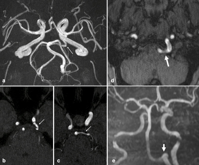
Figure 10.
Persistent vertebrobasilar anastomoses. (a) 3D MIP reconstruction and (b, c) axial TOF-MRA source images demonstrate a left-sided persistent trigeminal artery, emanating from the left cavernous ICA and anastomosing with the BA (small arrows). (d) Axial TOF-MRA and (e) 3D MIP reconstruction in another patient demonstrate a left-sided persistent hypoglossal artery, emanating from the left cervical ICA, traversing the hypoglossal canal (large arrows), and continuing as the basilar artery. The bilateral VAs were markedly hypoplastic. BA, basilar artery; ICA, internal carotid artery; MIP, maximum intensity projection; MRA, MR angiography; TOF, time-of-flight; VA, vertebral artery.

