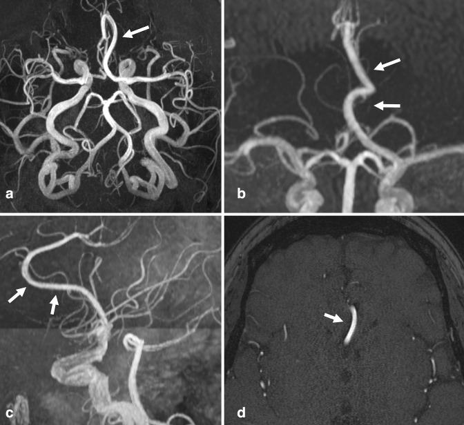
Figure 11.
Azygous anterior cerebral artery. (a) Axial oblique, (b) frontal, and (c) lateral views of a 3D MIP TOF-MRA, as well as (d) an axial TOF-MRA source image demonstrate an incidental single prominent midline ACA supplying the bilateral A3 branches (arrows). In this case, the large azygous artery is a continuation of the left A1 segment. A small right A1 segment supplied only a small orbitofrontal branch. MIP, maximum intensity projection; MRA, MR angiography; TOF, time-of-flight.

