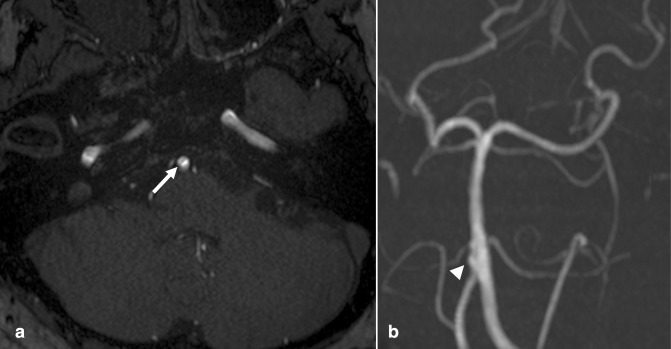
Figure 12.
Basilar artery fenestration. (a) Axial TOF MRA (b) frontal 3D MIP reconstruction demonstrate a linear luminal flow void at the base of the BA (arrow). Although appearance on axial images is similar to that of an intimal flap in the setting of dissection, and dilated appearance on 3D MIP (arrowhead) is similar to a dissecting aneurysm, location and short segment involvement are suggestive of a fenestration, a benign developmental variant. If findings on TOF MRA are equivocal, CTA or DSA can confirm. CTA, CT angiography; DSA, digital subtraction angiography; MIP, maximum intensity projection; MRA, MR angiography; TOF, time-of-flight.

