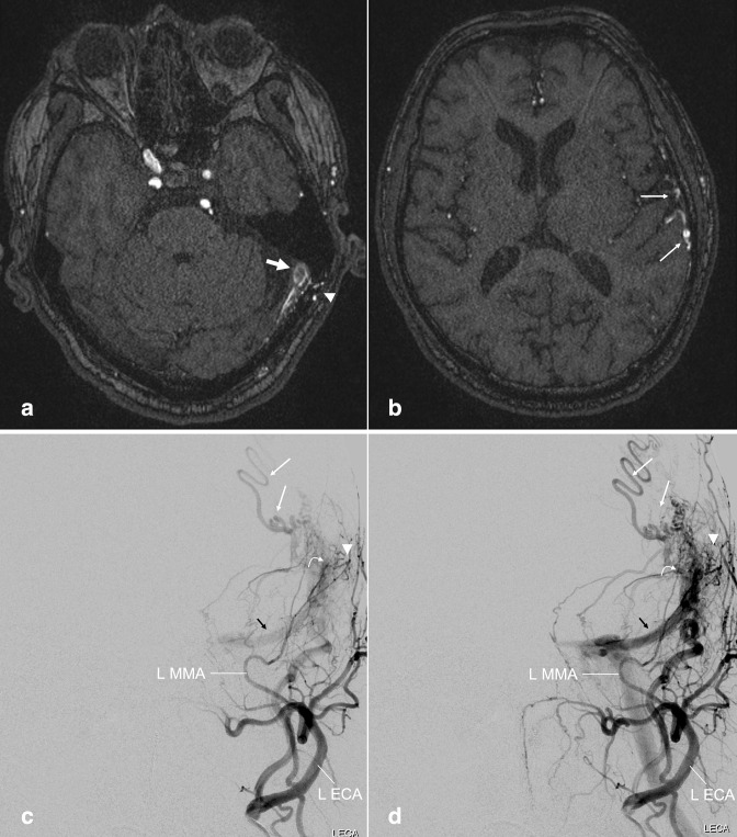
Figure 3.
Dural arteriovenous fistula. (a, b) Axial TOF MRA images performed on a patient with prior history of meningioma involving the superior sagittal sinus. Early flow-related enhancement in the dural venous sinus at the left transverse-sigmoid junction (a, large arrow), with adjacent transosseous venous collaterals (a, arrowhead), compatible with arteriovenous shunting. Prominent cortical veins with flow-related enhancement suggest venous backflow (b, small arrows). (c, d) Follow-up left external carotid artery injection DSA more fully demonstrates the dAVF, including the fistula nidus in which the middle meningeal artery branches empty directly into the transverse/sigmoid sinus (curved arrow), early filling of the left transverse sinus (black arrows), adjacent transosseous collaterals (arrowhead) and prominent cortical veins (small arrows). dAVF, dural arteriovenous fistula; DSA, digital subtraction angiography; MRA, MR angiography; TOF, time-of-flight.

