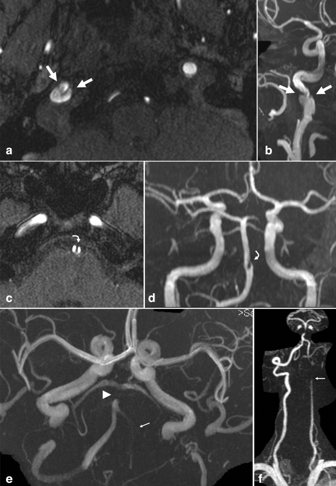
Figure 5.
Dissection. (a) Axial TOF-MRA and (b) 3D MIP reconstruction demonstrate an irregular, linear, luminal hypointensity with focal dilatation of the distal cervical ICA at the skull base (large arrows), representing intimal flap and pseudoaneurysm in the setting of dissection. (c) Axial TOF-MRA and (d) 3D MIP reconstruction from a different patient demonstrate an intimal flap of the BA with luminal irregularity (curved arrows). Subsequent VWI confirmed associated intramural hematoma, excluding a variant fenestration (not shown). (e) 3D MIP TOF-MRA in a different patient demonstrates an absent intracranial left VA (small arrow), and occlusion of the mid BA (arrowhead). (f) The contrast-enhanced neck MRA demonstrated tapering and occlusion of the left cervical VA, compatible with dissection (small arrow). BA, basilar artery; ICA, inferior cerebellar artery; MIP, maximum intensity projection; MRA, MR angiography; VA, vertebral artery; VWI, vessel wall MR imaging; TOF, time-of-flight.

