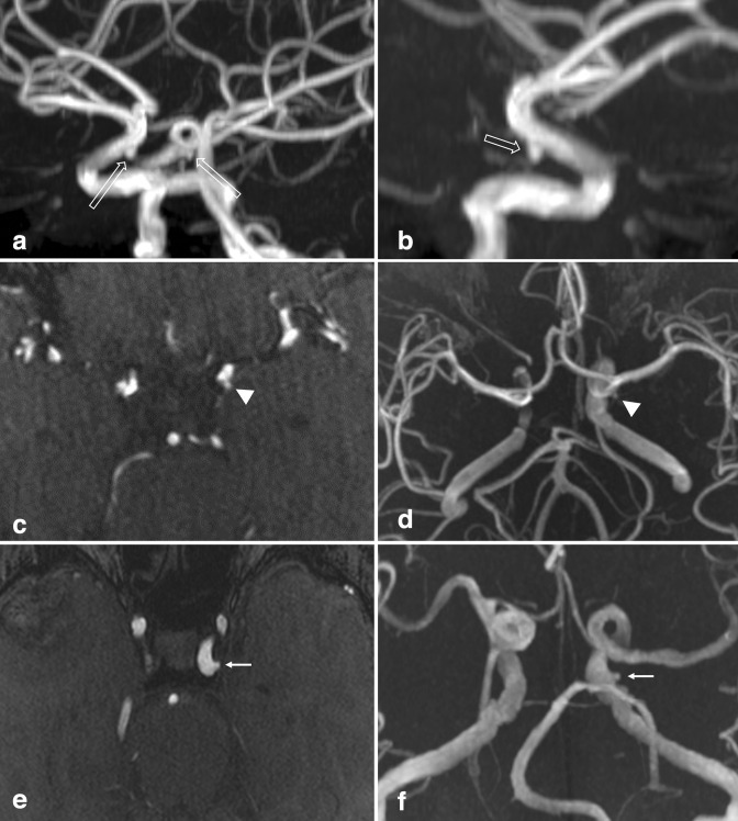
Figure 7.
Infundibulum. (a) Oblique view of a 3D MIP TOF-MRA of the brain demonstrates bilateral conical/triangular outpouchings projecting inferiorly from the terminal ICA (open arrows). Axial source images (not shown) clearly demonstrated small PCoAs emanated from the outpouchings, compatible with PCoA infundibula. (b) Lateral view of the right anterior circulation shows the 2 mm infundibulum and a faint PCoA (open arrow). (c) Axial TOF-MRA and (d) 3D MIP reconstruction demonstrate a small, 1–2 mm anterior choroidal artery infundibulum (arrowheads). (e) Laterally directed 1–2 mm outpouching from the cavernous ICA was favored to represent infundibulum (small arrows), given that a small vessel was seen emanating from the apex, likely the inferolateral trunk (not shown). ICA, inferior cerebellar artery; MIP, maximum intensity projection; MRA, MR angiography; PCoA, posterior communicating artery; TOF, time-of-flight.

