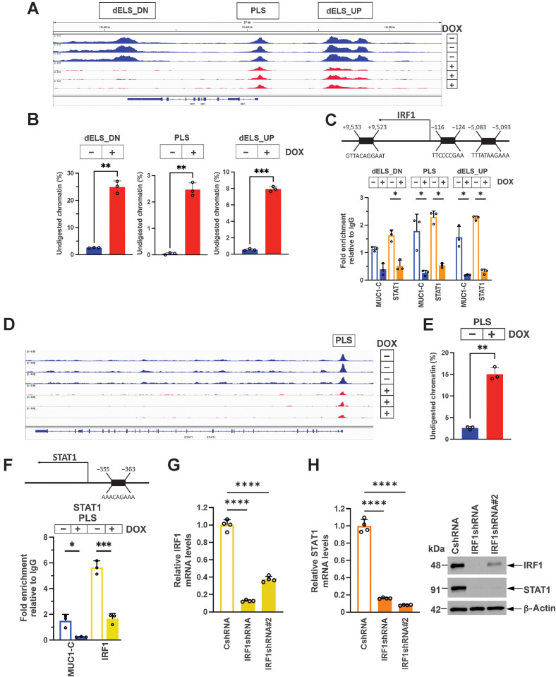
Figure 1.
MUC1-C drives chromatin accessibility and activation of IRF1 and STAT1. A, Genome browser snapshots of ATAC-seq data from the IRF1 gene in BT-549/tet-MUC1shRNA cells treated with vehicle or DOX for 7 days. B, Chromatin was analyzed for accessibility by nuclease digestion. The results are expressed as the percentage of undigested chromatin (mean±SD and individual values). C, Schema of the IRF1 gene highlighting positioning of a promoter-like signature (PLS) and distal enhancer-like signatures (dELS) downstream (dELS-DN) and upstream (dELS-UP) to the TSS. Soluble chromatin from BT-549/tet-MUC1shRNA cells treated with vehicle or DOX for 7 days was precipitated with a control IgG, anti–MUC1-C and anti-STAT1. The DNA samples were amplified by qPCR with primers for the indicated IRF1 regions. The results (mean ± SD and individual values) are expressed as fold-enrichment as compared with that obtained from control IgG-precipitated chromatin (assigned a value of 1). D and E, Genome browser snapshot of ATAC-seq data from the STAT1 PLS region in BT-549/tet-MUC1shRNA cells treated with vehicle or DOX for 7 days (D). Chromatin was analyzed for accessibility by nuclease digestion (E). The results are expressed as the percentage of undigested chromatin (mean ± SD and individual values). F, Schema of the STAT1 gene with localization of a PLS upstream to the TSS. Soluble chromatin from BT-549/tet-MUC1shRNA cells treated with vehicle or DOX for 7 days was precipitated with a control IgG, anti–MUC1-C and anti-IRF1. The DNA samples were amplified by qPCR with primers for the STAT1 PLS region. The results (mean±SD and individual values) are expressed as relative fold enrichment as compared with that obtained with IgG (assigned a value of 1). G, BT-549/CshRNA, BT-549/IRF1shRNA and BT-549/IRF1shRNA#2 cells were analyzed for IRF1 and STAT1 mRNA levels by qRT-PCR. The results (mean ± SD and individual values) are expressed as relative mRNA levels as compared with that obtained in CshRNA cells (assigned a value of 1). H, Lysates were immunoblotted with antibodies against the indicated proteins. *, P ≤ 0.05; **, P ≤ 0.01; ***, P ≤ 0.001; ****, P ≤ 0.0001.

