
Figure 3. Polyglutamylation controls the microtubule‐severing activity of katanin.
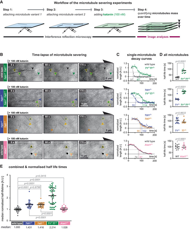
- Schematic representation of the katanin severing assay. Label‐free Taxol‐stabilised microtubules, polymerised from wild‐type and knockout‐mouse brain tubulin, are attached to a glass coverslip sequentially and imaged in IRM mode. Their identity is retained by their positions at the coverslip (Steps 1 and 2). Next, 100 nM purified p60/p80 katanin complex is added to the chamber (Step 3). The change in the microtubule length over time is recorded in IRM and quantified in subsequent analyses (Step 4).
- Representative IRM still images at different time points from time‐lapse following the addition of 100 nM p60/p80 katanin complex on microtubules (Movie EV3). Arrowheads indicate microtubules of a certain type (colour‐coded, as labels on the left). Scale bars 5 μm.
- Decay curves of normalised microtubule length plotted against time upon incubation with katanin (A.U., arbitrary units) for single microtubules highlighted by arrow heads with yellow borders in (B).
- Half‐life times (seconds) of all microtubules from the assays of which representative images are displayed in (B) and representative decay curves are shown in (C). Each data point represents the half‐life of a single microtubule. Medians with interquartile ranges are shown. Mann–Whitney test, P‐values displayed.
- Analysis of normalised microtubule half‐lifetimes from 14 independent experiments (Fig EV3). Each data point represents half‐life of a single microtubule. Medians with interquartile ranges shown, Mann–Whitney test and P‐values displayed.