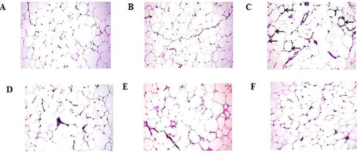
Fig. 3.

Photomicrograph of the visceral adipose tissues stained with hematoxylin and eosin of (A) control rats shows normal white adipose tissue with large round adipocytes, (B) oleic acid group showing normal adipose tissue, (C) HFD group showing numerous necrotic adipocytes surrounded by macrophages forming distinctive crown-like structure (arrows), (D) HFD-exercise group showing mild leukocytic cell infiltration, (E) HFD-oleic acid group showing large adipocytes, and (F) HFD-exercise-oleic acid group showing minimal macrophages and lymphocytic cell infiltration; magnification 20×. HFD, High-fat diet.
