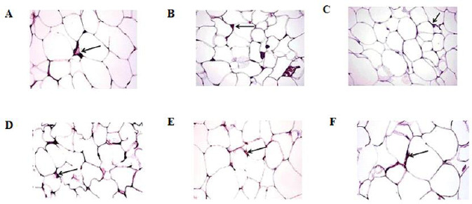
Fig. 6.

photomicrograph of the visceral adipose tissues stained for CD206 shows positive cells at the junction of two or more adipocytes (arrow) of (A) control group, (B) oleic acid group, (C) HFD group, (D) HFD-exercise group, (E) HFD-oleic acid group, and (F) HFD-exercise-oleic acid group. (CD206immunohistochemical staining, 40×. HFD, High fat diet.
