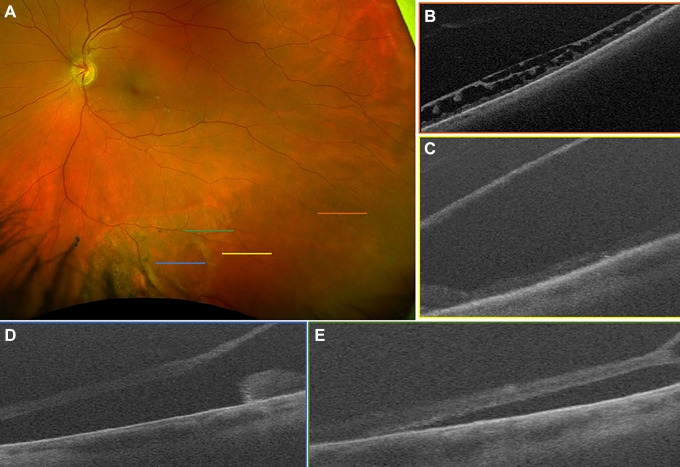
Figure 1.
(A) Scanning laser ophthalmoscope image demonstrating inferior retinoschisis with large cavity, outer retinal break, and associated retinal detachment. No inner retinal break was identified on examination. (B) Shallow retinoschisis, located temporally and correlating with the orange raster marker in (A), was noted on ultra-widefield swept-source optical coherence tomography. (C) A more bullous retinoschisis cavity was clearly demonstrated to correlate with the yellow raster marker in (A). (D) An outer retinal break with rolled margins was captured with the blue raster marker in (A), with (E) subretinal fluid nasal and just posterior (green raster marker in [A]) to the retinoschisis cavity. The patient has been observed without progression for 6 months.

