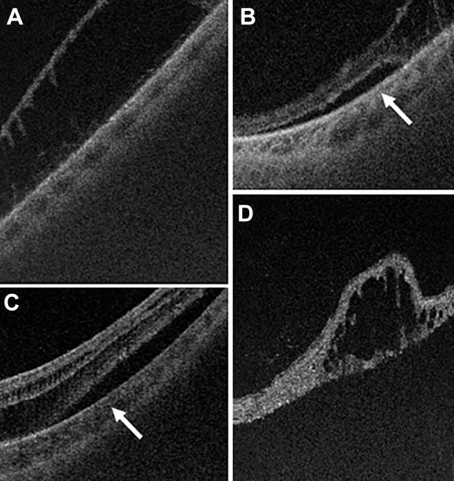
Figure 2.

(A) Ultra-widefield swept-source optical coherence tomography (UWF SS-OCT) images used to confirm the presence of asymptomatic retinoschisis. (B) Occasionally, subretinal fluid cuffs adjacent to an area of retinoschisis can be diagnosed and monitored with serial UWF SS-OCT images. Patients in both (A) and (B) were asymptomatic. (C) The appearance was distinct from subretinal fluid (arrow) in combined retinoschisis and retinal detachment in a symptomatic patient with acute peripheral vision loss. (D) Degenerative retinal cyst in an area of chronically detached retina can sometimes clinically be mistaken for retinoschisis; however, it can definitively be identified with UWF SS-OCT.
