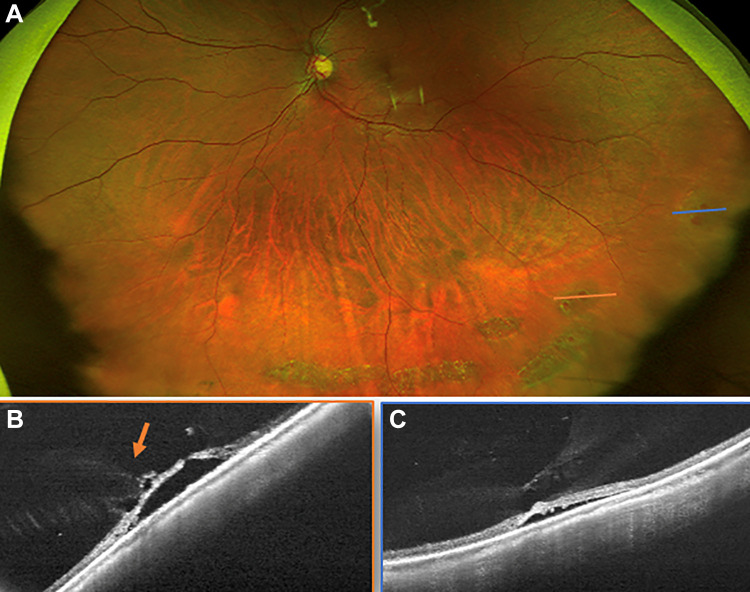
Figure 3.
(A) A scanning laser ophthalmoscope image demonstrating inferior lattice and retinal holes. The orange line shows the location of the (B) ultra-widefield swept-source optical coherence tomography raster through an atrophic hole, which confirmed the hole had subretinal fluid and vitreous traction (orange arrow). The blue line in (A) shows the area of (C) an ultra-widefield swept-source optical coherence tomography through a temporal retinal hole, which also confirmed vitreous traction with associated subretinal fluid. The decision to perform prophylactic laser barrier retinopexy was made based on the clinical appearance of holes with fluid in a patient with symptoms and was supported by the findings that confirmed vitreous traction on the holes’ margins on imaging.

