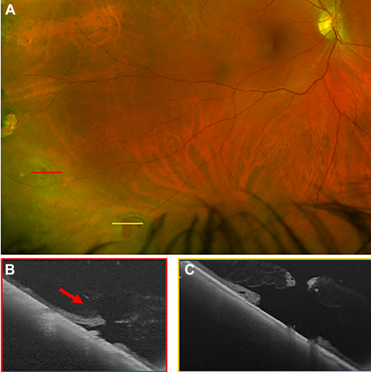
Figure 4.
(A) A scanning laser ophthalmoscope image with inferotemporal retinal breaks. The red line shows the location of (B) an ultra-widefield swept-source optical coherence tomography raster through the superior tear, which had vitreous traction (red arrow). The yellow line in (A) shows the location of (C) an ultra-widefield swept-source optical coherence tomography raster through an inferior retinal break that had a flat retina without flap, subretinal fluid, or vitreous traction. Imaging supported the clinical decision for treatment of the symptomatic retinal break with persistent vitreous traction. In this case the second break was also treated at the same time because laser was already being performed and the patient was acutely symptomatic.

