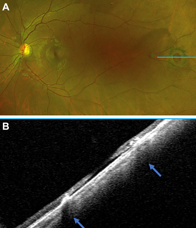
Figure 5.

(A) A scanning laser ophthalmoscope image showing an old, scarred down, pigmented retinal hole with an optical coherence tomography scan through the blue line. (B) An ultra-widefield swept-source optical coherence tomography scan demonstrating a flat retinal hole with no traction and scarred down edges (arrows). Supported by the imaging findings, the patient was observed without intervention.
