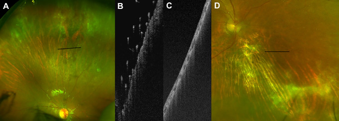
Figure 6.
(A) Color image of retinal periphery demonstrating an active cytomegalovirus lesion. (B) Ultra-widefield swept-source optical coherence tomography of lesion, with preretinal hyperreflective foci suggestive of disease activity. (C) Ultra-widefield swept-source optical coherence tomography and (D) color images of a different patient with an old, inactive cytomegalovirus lesion that had no preretinal hyperreflective foci, suggesting absence of activity.

