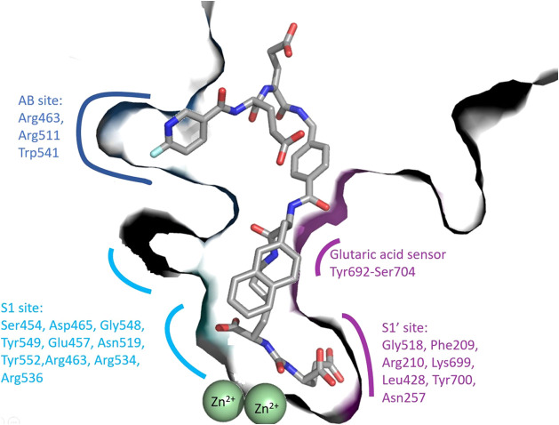
Figure 1.
Structure of the GCPII/PSMA-1007 complex (PDB entry 5o5t 21) illustrates the anatomy of the GCPII internal cavity and the S1, S1′, and AB site (ABS) pockets. Zinc atoms are shown as green spheres. The S1 binding site consists of residues Ser454, Asp465, Gly548, Tyr549, Glu457, Asn519, Tyr552, and a positively charged arginine patch (Arg463, Arg534, and Arg536, pale cyan). The S1′-binding site (purple) is formed by residues Gly518, Phe209, Arg210, Lys699, Leu428, Tyr700, and Asn257. Residues 692–704 constitute a glutaric acid sensor. The arene binding site (ABS blue) is formed by Arg463, Arg511, and Trp541.

