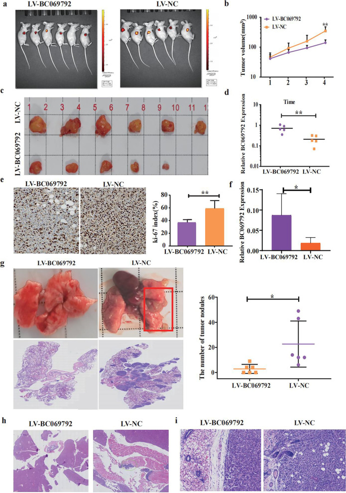
Fig. 3.
BC069792 inhibits the proliferation and invasion of breast cancer cells in vivo. a In vivo imaging of nude mice showed that the volume of the subcutaneous tumor in the LV-BC069792 group was significantly smaller than that of the LV-NC group. One nude mouse in the LV-BC069792 group had no tumor. b The growth curve showed that the tumor growth rate of the LV-NC group was significantly higher than that of the LV-BC069792 group (P = 0.0081). c The removal of subcutaneous tumors showed that the tumor volume in the LV-BC069792 group was significantly smaller than that of the LV-NC group, and the tumor of a nude mouse in the LV-BC069792 group were suppressed. d The content of BC069792 in tumor tissue in LV-BC069792 group was significantly higher than that in LV-NC group (P = 0.0051). e The Ki-67 index of the LV-BC069792 group was about 25%, and the Ki-67 index of the LV-NC group was about 80% (IHC × 200). The Ki-67 index of the LV-BC069792 group was significantly lower than that of the LV-NC group (P = 0.0059). f In lung metastasis, the expression of BC069792 in LV-BC069792 group was significantly higher than that in LV-NC group (P = 0.0228). g The gross image of the lung metastases in nude mice showed that the tumor growth at the hilum and outside the lung of the nude mice in the LV-NC group was obvious. In LV-BC069792 group, there were no obvious tumors in the lungs of nude mice. HE staining results showed that the lung metastases in the LV-NC group were significantly increased and the volume was significantly increased. The scatter plot showed that the number of metastases in the overexpression BC069792 group was significantly reduced (P = 0.0267). h There was liver-diaphragm metastasis in the LV-NC group, but no cancer metastasis in the liver and diaphragm of the LV-BC069792 group. i The tumor cells in the LV-NC group invaded the skin adnexa, while the tumor cells in the LV-BC069792 group had a clear boundary with the surrounding adipose tissue and mammary tubules, and no obvious invasion was found. (P < 0.05, P < 0.01, P < 0.001,.P < 0.0001, ns represent P > 0.05)**********

