Figure 1:
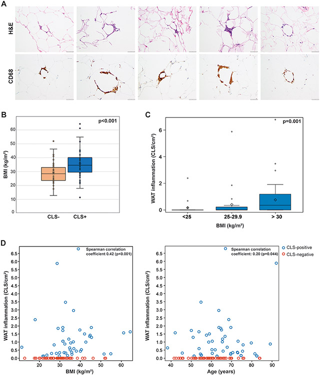
(A) Crown-like structures (CLSs) in endometrial cancer. Hematoxylin and eosin–stained sections of adipose tissue derived from patients with newly diagnosed endometrial cancer (left), and results of anti-CD68 immunohistochemical analysis of macrophages identified CLSs (right). (B) Box plot displaying the distribution of body mass index (BMI) based on the presence or absence of CLS/white adipose tissue (WAT) inflammation in patients with endometrial cancer. CLS-positive patients had a median BMI of 34.6 kg/m2 (range, 11.4-64.3), compared to 28.5 kg/m2 (range, 12.8-52.3) for CLS-negative patients (p<0.001). Wilcoxon rank sum test was performed. (C) Box plot displaying the quantity of WAT inflammation by BMI category among all patients. The median quantity of WAT inflammation was 0.00 CLS/cm2 (range, 0.00-2.39) for patients with a BMI <25 kg/m2, 0.00 CLS/cm2 (range, 0.00-5.88) for patients with a BMI of 25-29 kg/m2, and 0.35 CLS/cm2 (range, 0.00-6.79) for patients with a BMI ≥30 kg/m2. Kruskal-Wallis test was performed. (D) Scatterplots demonstrating measures of association between levels of WAT inflammation and BMI or age as continuous variables. Spearman rank correlation coefficients (nonparametric measures of association) were reported.

