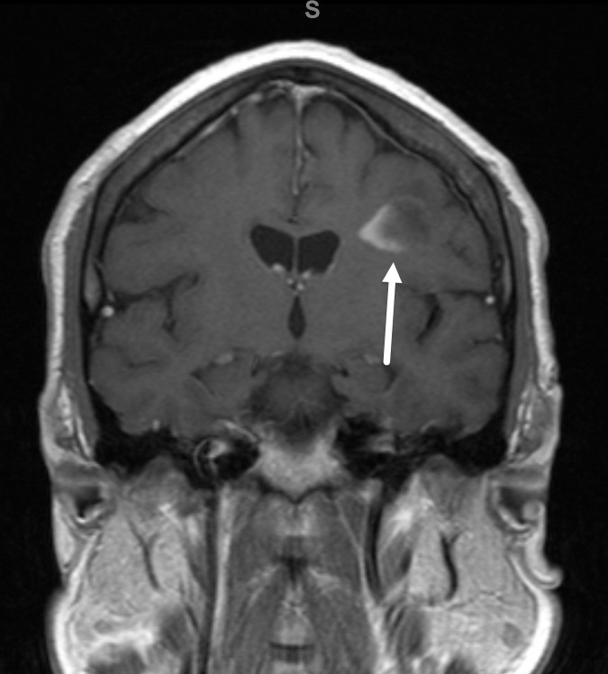
Figure 3.
Incomplete rim enhancement of the left frontal lobe lesion. Post-contrast coronal T1 MR image through the lesion in Figure 2 demonstrates incomplete rim enhancement open to the cortical surface (arrow). This finding is typical of demyelination.

