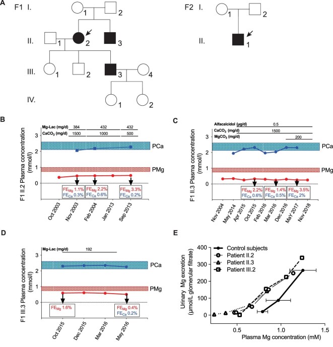
FIGURE 1:
HSH phenotype in two families with a dominant inheritance pattern. (A) Pedigree of the families with autosomal dominant hypomagnesaemia and secondary hypocalcaemia. Black symbols denote affected and genetically confirmed family members. (B–D) Clinical course of patients F1-II.2, II.3 and III.3. (E) Mg2+ loading test showing combined intestinal and renal Mg2+ wasting in individuals F1-II.2, F1-II.3 and F1-III.3. In patients on baseline (fasting) conditions, plasma Mg concentration was low and urinary Mg excretion was low as well (7.4 ± 6.6 µmol/L of glomerular filtrate), as expected under fasting conditions and in the same range as in control subjects under control conditions (19.2 ± 1.9 µmol/L of glomerular filtrate), showing that no urinary loss of Mg is seen when plasma Mg concentration is low. Then plasma Mg increased during Mg infusion in patients and controls and urinary Mg increased as well. However, for any plasma Mg concentration, urinary Mg excretion was higher in patients than in controls, showing that renal tubular reabsorption is impaired in patients; this can be clearly seen for any plasma Mg concentration ≥0.8 mmol/L. Therefore we conclude that the defect in renal tubular Mg reabsorption in patients manifests when plasma Mg reabsorption is normal or high.

