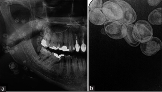
Figure 2.

Radiographic aspects (a) Cropped panoramic radiograph demonstrates multiple, round well-defined radiopaque lesions with radiolucent edges of variable sizes along buccal region, extending from coronoid process region up to the base of the mandible, (b) Periapical radiograph of right buccal mucosa demonstrating multiple radiopaque lesions with radiolucent edges, variable sizes, rounded and oval shapes
