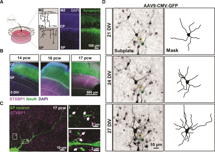
Figure 1.
Human organotypic cortical brain slices cultures with developing subplate neurons. (A) Schematic and low magnification photomicrographs of organotypic cultures prepared from human foetal brain tissue (created with BioRender.com). Cultures are maintained for 1–4 weeks following infection with and without different viral vectors. Micrograph image represents a 16 pcw sample, 2 weeks in culture with clearly identifiable cortical regions including the marginal zone (MZ), cortical plate (CP) and subplate (SP). DAPI labels all cell nuclei and Synapsin1 immunohistochemistry labels reveals pre-synaptic terminals predominantly located in SP but also CP and MZ. (B) Representative images from 14, 16 and 17 pcw cultures (0 DIV). STXBP1 and NeuN (all neurons) immunofluorescence reveals location of the SP. Note the enrichment of STXBP1 within the SP at 16–17 pcw (n = 3 human samples/age). (C) SP neuron (eGFP transduced cell) with STXBP1 present on neurites along thin putative axons extending from the cell body [C(i)] and spine-like structures [C(ii)] indicated by the arrows. (D) Photomicrographs of a 16 pcw culture infected with AAV9-CMV-GFP and taken at 7, 21 and 27 DIV, showing the same neuron (highlighted by the masked images). Note the dynamic nature and elaboration of neurites over time. Coloured outlines highlight reference cells that are stable overtime.

