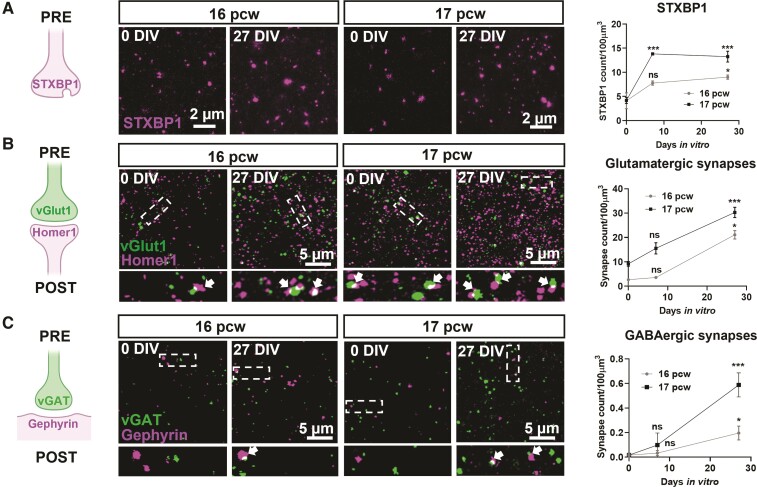
Figure 2.
Synapse maturation in human cortical slice cultures. (A) Left shows the schematic displaying the pre-synaptic location of STXBP1. Middle shows the photomicrographs of STXBP1 in the subplate of 16–17 pcw cultures at 0 and 27 DIV. Right shows the quantification of the increase in the number of STXBP1 puncta over time at both ages. (B) Left to middle shows the colocalization of the pre- and post-synaptic glutamatergic markers, vGlut1 and Homer1, respectively indicated by arrows, in the subplate at 0 and 27 DIV. Areas, demarcated by dotted boxes in the upper micrographs, are shown enlarged below. All cultures were prepared at 16–17 pcw. (C) Similar analyses performed for GABAergic synapses based on colocalization of vGAT (pre-synaptic) and Gephyrin (post-synaptic). Analyses for A–C were performed as two-way ANOVA with repeated measures from comparisons made at 0 DIV for each age, n = 6–8 slices per time point from 2–3 human samples/age. Error bars indicate SEM.

