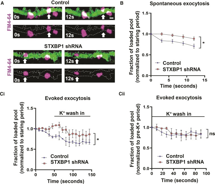
Figure 4.
Impaired vesicle exocytosis with reduced STXBP1 levels. (A) Isolated axons (green) tracked from FM4-64 loaded subplate neurons in scrambled control and STXBP1 shRNA slices (16 pcw) at 14 DIV. Note the presence of spontaneous exocytosis from vesicles loaded with FM4-64 (arrows). Scale bar = 1 µm. (B) Quantification of spontaneous exocytosis (n = 12 slices from four human samples) in both genetic conditions (without stimulation). (C) Quantification of evoked exocytosis in separate slices (n = 8 slices from three human samples) following brief potassium chloride (K+) stimulation. Data were normalized to the first five frames of the recording (starting period) [C(i)] or to the five frames immediately prior to K+ wash in (pre-K+ period) [C(ii)]. All analysis was performed using a two-way ANOVA with repeated measures. All error bars represented as SEM.

