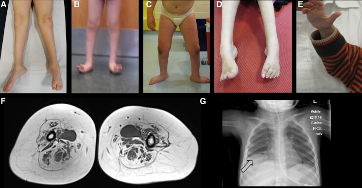
Figure 3.
Clinical and radiological features of neuronopathies. (A) Distal leg thinning in a patient with MFN2 variant; (B) lower leg atrophy and foot deformities in a patient with BICD2 variant; (C) lower leg atrophy and foot posture in a patient with DYNC1H1 variant. (D) Foot posture in a young adult with TRPV4 variant; (E) photo of hand atrophy (split-hand sign) in a patient with GARS variant; (F) thigh muscle MRI showing islands of muscle, a finding strongly suggestive of neuronopathy; (G) X-ray showing diaphragmatic paralysis in SMARD1 (arrow).

