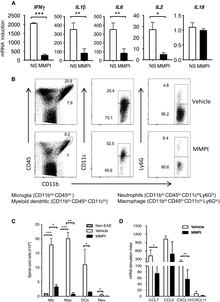
Fig. 7. MMPt-induced T cell depletion leads to suppression of inflammatory cytokines in the CNS of MOGp-induced C57BL/6 EAE mice.
(A) Quantitative PCR results for indicated cytokines from the spinal cord mononuclear cells isolated from mice of day 20 post-EAE induction that were treated at day 14 of disease scoring of 3 with IV injections of one dose of 800 μg of MMPt or normal saline (NS) control. (B) Frequency of microglia (Mic), myeloid DCs, macrophages (Mac), and neutrophils (Neu) in the affected CNS of mice that received three doses of vehicle control or 800 μg of MMPt every other day starting from day 14 of EAE induction that presented with a disease score of 3 is shown with gating, and each specific population is specified at the bottom. (C) Quantification of (B) with absolute cell counts on average of three mice per group. (D) mRNA stimulation index derived from quantitative PCR data measuring expression of chemokines of EAE CNS tissue isolated from EAE mice that received one dose of IV injection of normal saline or 800 μg of MMPt for 20 hours. Data were analyzed using two-tailed unpaired t test (shown means + SD); *P < 0.05, **P < 0.01, and ***P < 0.001. Data represent two independent experiments.

