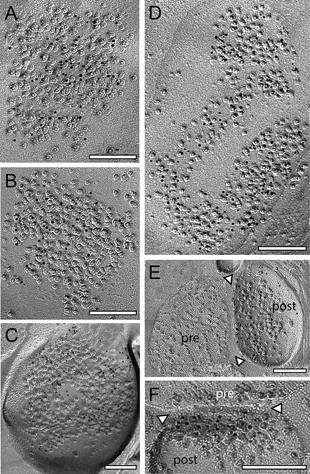
Fig. 3.

Density and distribution patterns of AMPA receptors and the GluN1-subunit of the NMDA-receptor at dendritic shaft and spine PSDs at L4 synaptic contacts. A–B) Two examples of 2 large shaft PSDs with a macular, non-perforated appearance labeled with gold grains detecting for AMPA- A) and the GluN1 B). C) Ring-like spine PSD labeled with gold grains detecting the GluN1. D) Large dendritic shaft PSD with a horseshoe to ring-like appearance labeled with gold grains detecting AMPA receptors. E, F) Two spine PSDs with a comparably low density of gold grains detecting AMPA E) and the GluN1 F). In both images, the synaptic cleft is marked by arrowheads. Abbreviations: pre: presynaptic; post: postsynaptic. Scale bars in A–F) 0.1 μm.
