Figure 2.
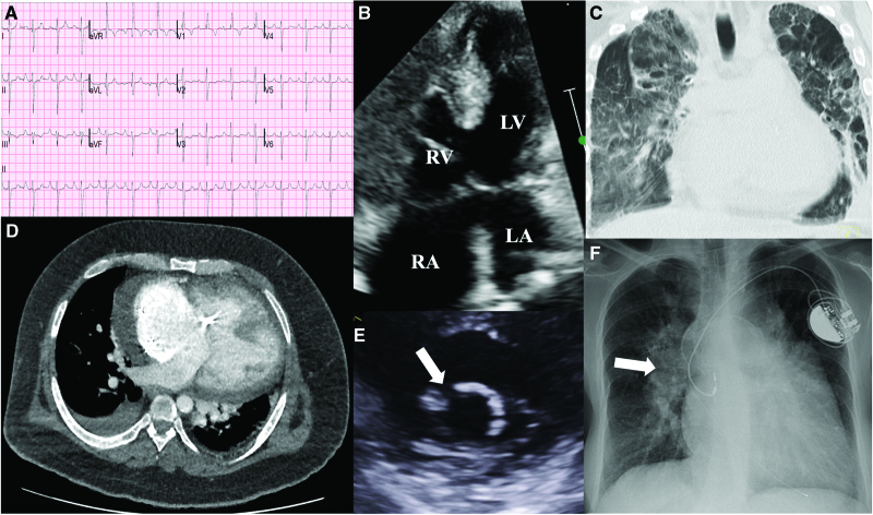
Manifestations of congenital heart disease in Down syndrome. A, ECG of an individual with Down syndrome, atrioventricular septal defect (AVSD), and Eisenmenger syndrome. There is right bundle branch block, peaked P waves (P pulmonale), and extreme QRS axis. B, A complete AVSD is shown with low velocity bidirectional shunting at atrial and ventricular levels. C, Computed tomography scan of the thorax (coronal section) in a person with Eisenmenger ventricular septal defect, displaying gross cardiomegaly along with severe bronchopulmonary dysplasia. D, Axial computed tomography image from an individual with Down syndrome, obesity, and Eisenmenger syndrome with complete AVSD and a permanent pacemaker. E, Parasternal short-axis view of a trileaflet left atrioventricular valve after AVSD repair. The arrow shows the gap between the 2 bridging leaflets, which is commonly the site of regurgitation. F, Chest radiography shows a dual chamber permanent pacemaker in an individual with Down syndrome and Eisenmenger AVSD. There is severe dilation of the pulmonary vasculature, most visible on the right (arrow), and severe cardiomegaly. LA indicates left atrium; LV, left ventricle; RA, right atrium; and RV, right ventricle.

