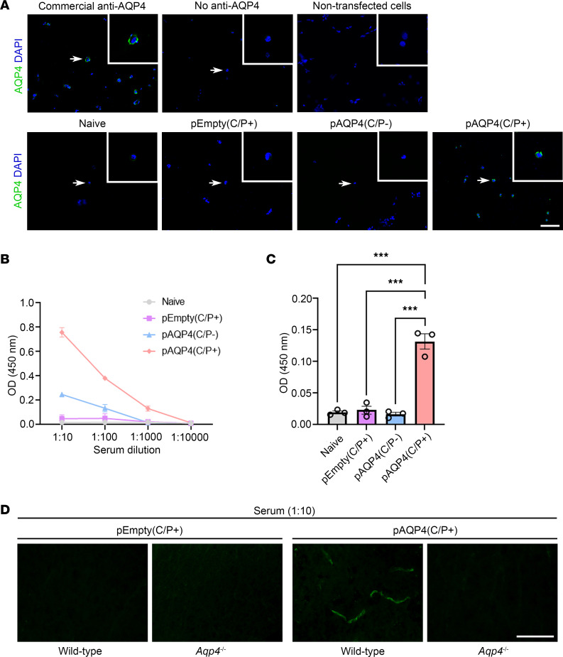
Figure 2. AQP4 immunization generates AQP4 autoantibodies.
(A) Detection of AQP4 autoantibodies in mouse serum by cell-based indirect immunofluorescence assay. HEK293 cells were transfected with a plasmid encoding the mouse AQP4 M23 isoform. AQP4 autoantibodies in the serum were visualized by fluorescence-conjugated secondary antibody specific for mouse IgG. Top left: Immunostaining with commercial anti-AQP4 antibody revealed a discontinuous pattern of AQP4 staining on the cell membrane (positive control). Top middle and right: No AQP4 immunoreactivity was observed when commercial anti-AQP4 antibody was absent or HEK293 cells were not transfected (negative controls). Bottom: Immunostaining of transfected HEK293 cells using the serum of naive, pEmpty(C/P+), pAQP4(C/P–), and pAQP4(C/P+) mice. Nuclei were counterstained with DAPI. Images are representative of 8 mice per group. Scale bar: 50 μm. Original magnification, ×400 (insets). (B) Titer of AQP4 autoantibodies was measured by ELISA using serial dilution of serum from 1:10 to 1:10,000. (C) Concentration of AQP4 autoantibodies was determined using serum diluted at 1:1,000. (D) Spinal cord sections of WT and Aqp4-deficient (Aqp4–/–) mice were immunostained using the serum of pEmpty(C/P+) and pAQP4(C/P+) mice. Images are representative of 3 mice per group. Data are mean ± SEM; n = 3 per group. ***P < 0.001, 1-way ANOVA with post hoc Tukey’s test. Scale bar: 50 μm.

