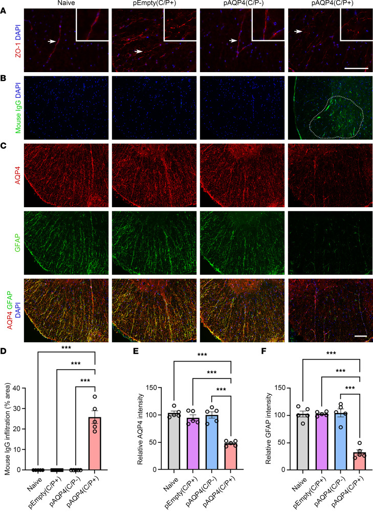
Figure 4. IgG infiltration and astrocytopathy after AQP4 immunization.
(A) Immunostaining for ZO-1 in the spinal cord of naive, pEmpty(C/P+), pAQP4(C/P–), and pAQP4(C/P+) mice. Insets are higher-magnification photomicrographs showing the pattern of ZO-1 staining in blood vessels. Original magnification, ×400 (insets). (B) Immunostaining for mouse IgG in the spinal cord of naive, pEmpty(C/P+), pAQP4(C/P–), and pAQP4(C/P+) mice. Dotted line demarcates the area of mouse IgG immunoreactivity. (C) Coimmunostaining for AQP4 and GFAP in the spinal cord of naive, pEmpty(C/P+), pAQP4(C/P–), and pAQP4(C/P+) mice. (D) Quantification of mouse IgG infiltration. (E and F) Quantification of AQP4 and GFAP immunofluorescence intensities. Images are representative photomicrographs showing the ventrolateral white matter of cervical spinal cord cross sections from 5 mice per group. Nuclei were counterstained with DAPI. Data are mean ± SEM; n = 5 per group. ***P < 0.001, 1-way ANOVA with post hoc Tukey’s test. Scale bars: 50 μm.

