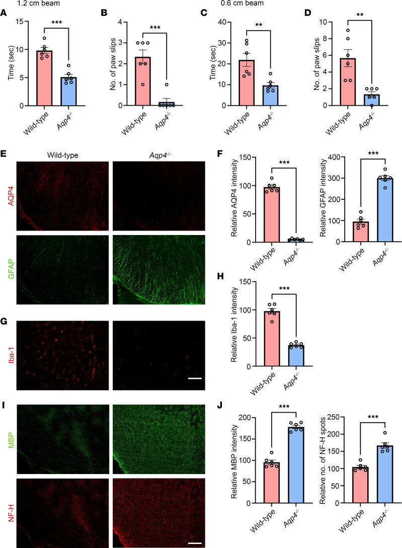
Figure 7. AQP4 immunization does not induce motor impairments and spinal cord pathologies in Aqp4-deficient mice.
WT or Aqp4-deficient (Aqp4–/–) mice received in vivo electroporation of pAQP4 at the left tibialis anterior muscle. Electroporation was performed at days 0, 14, and 28. Animals were culled at day 42. (A and B) Beam walking test measuring time taken by a mouse to cross and number of paw slips while crossing a 1.2 × 80 cm (width × length) beam. (C and D) Time taken to cross and number of paw slips while crossing a 0.6 × 80 cm beam. (E) Immunostaining for AQP4 and GFAP in the spinal cord of WT pAQP4(C/P+) and Aqp4–/– pAQP4(C/P+) mice. (F) Quantification of AQP4 and GFAP immunofluorescence intensities. (G) Immunostaining for Iba-1 in the spinal cord of WT pAQP4(C/P+) and Aqp4–/– pAQP4(C/P+) mice. (H) Quantification of Iba-1 immunofluorescence intensity. (I) Immunostaining for MBP and NF-H in the spinal cord of WT pAQP4(C/P+) and Aqp4–/– pAQP4(C/P+) mice. (J) Quantification of MBP immunofluorescence intensity and number of NF-H spots. Images are representative photomicrographs showing the ventrolateral white matter of cervical spinal cord cross sections from 6 mice per group. Data are mean ± SEM; n = 6 per group. **P < 0.01, ***P < 0.001, Student’s 2-tailed t test. Scale bars: 50 μm.

