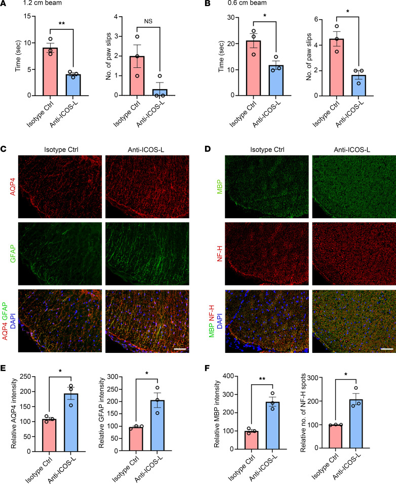
Figure 9. Tfh cell depletion ameliorates NMOSD disease activity.
Beginning at day 0, pAQP4(C/P+) mice were given 150 μg anti–ICOS-L or isotype control antibody (i.p.) 3 times a week. Animals were culled at day 42. (A and B) Beam walking test measuring time taken by a mouse to cross and number of paw slips while crossing a 1.2 × 80 cm beam (A) and a 0.6 × 80 cm beam (B). (C) Coimmunostaining for AQP4 and GFAP in the spinal cord of pAQP4(C/P+) mice following isotype control or anti–ICOS-L treatment. (D) Coimmunostaining for MBP and NF-H in the spinal cord of pAQP4(C/P+) mice following isotype control or anti–ICOS-L treatment. (E) Quantification of AQP4 and GFAP immunofluorescence intensities. (F) Quantification of MBP immunofluorescence intensity and number of NF-H spots. Images are representative photomicrographs showing the ventrolateral white matter of cervical spinal cord cross sections from 3 mice per group. Nuclei were counterstained with DAPI. Data are mean ± SEM; n = 3 per group. *P < 0.05, **P < 0.01, Student’s 2-tailed t test. Scale bars: 50 μm.

