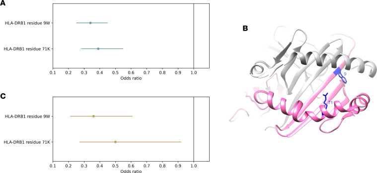
Figure 3. Association of ADA development with HLA-DRB1 residues 9 and 71 within the peptide-binding groove.
(A) Estimated odds ratios for the presence of a tryptophan residue at position 9 (9W) and a lysine residue at position 71 (71K) of HLA-DRB1 on risk of ADA development in the discovery cohort (individuals sampled 6–36 months after treatment initiation, n = 784). Effect sizes and 95% confidence intervals estimated jointly for both variants in a multiple regression model. (B) Three-dimensional ribbon model of the HLA-DR protein. Structure based on Protein Data Bank entry 3pdo, with a direct view of the peptide-binding groove. HLA-DRB is shown in pink; HLA-DRA is shown in gray. The 2 key amino acid positions identified by the association analyses are shown with their side chains and highlighted in blue. (C) Estimated odds ratios for the presence of a tryptophan residue at position 9 (9W) and a lysine residue at position 71 (71K) of HLA-DRB1 on risk of ADA development in the replication cohort (individuals sampled within 6 months of treatment initiation, n = 232). Effect sizes and 95% confidence intervals estimated jointly for both variants in a multiple regression model.

