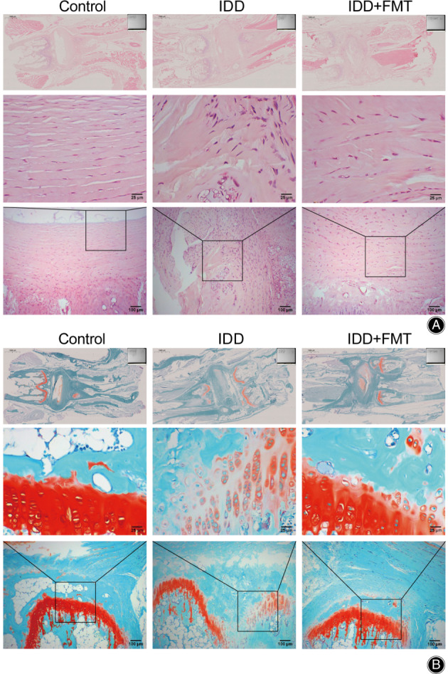
Fig. 1.

Histopathological analysis of vertebral disc after FMT. (A) A micrograph showing HE staining of rat intervertebral disc tissue. (Scale bar = 25/100/1000 μm). (B) A micrograph showing S‐O staining of rat intervertebral disc tissue. (Scale bar = 25/100/1000 μm). FMT, fecal microbiota transplantation.
