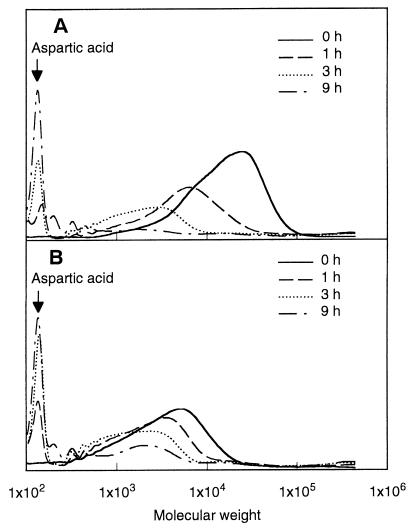
FIG. 3.
Time-dependent changes in the molecular weights of PAA-P (A) and PAA-T (B) treated with cell extract of strain KT-1. Cells of strain KT-1 were disrupted by ultrasonic treatment, and the cell extract was incubated with 0.15% (wt/vol) PAA in carbonate buffer (pH 7.0) at 28°C. The solutions were then removed and analyzed by GPC.

