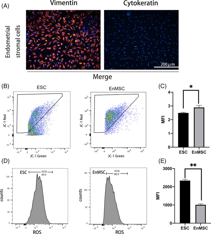
FIGURE 2.

Endometrial mesenchymal stem cells (EnMSCs) displayed higher mitochondrial membrane potential (ΔΨm) and lower reactive oxygen species (ROS) than primary endometrial stromal cells (ESCs). (A) Identification of ESCs by immunofluorescence staining. Primary ESCs were positive for vimentin expression and negative for cytokeratin expression. Bar = 200 μm. (B) Flow cytometry analysis was carried out to detect the △Ψm in primary ESCs and EnMSCs by JC‐1 staining. (C) Red/green fluorescence intensity was calculated in both kinds of cells, and EnMSCs had a higher △Ψm than primary ESCs. (D) ROS levels were measured by DCFH‐DA fluorescence in both cell lines via flow cytometry. (E) A reduced ROS fluorescence intensity was found in primary ESCs. In (C) and (E), the data are the mean ± SEM of three replicates. One asterisk (*) indicates statistically significant differences at p < 0.05, and two asterisks (**) indicate statically significant differences at p < 0.01.
