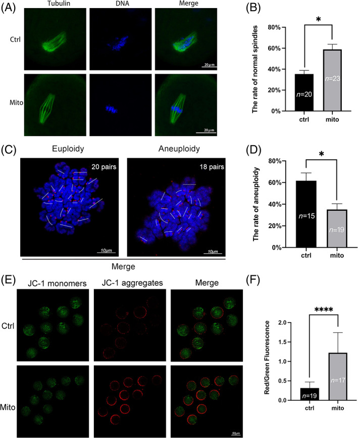
FIGURE 4.

The quality of oocytes from aged mice was improved by endometrial mesenchymal stem cell (EnMSC) mitochondrial transfer. (A) Representative pictures of the spindles and chromosomes in the two groups. (B) Increased normal spindle rates were observed in MII oocytes from aged mice after mitochondrial supplementation. Bar = 20 μm. (C) Images of chromosome spreads are shown. Bar = 10 μm. (D) Decreased aneuploidy rates were found in MII oocytes after mitochondrial transfer. (E) The mitochondrial membrane potential (△Ψm) values of the two groups were assessed by JC‐1 staining in MII oocytes after mitochondrial supplementation. JC‐1 monomers with green fluorescence revealed a low △Ψm, while JC‐1 aggregates with red fluorescence revealed a high △Ψm. Bar = 50 μm. (F) Increased △Ψm was observed in MII oocytes from aged mice after mitochondrial supplementation. The data are the mean ± SEM. In C, D and F, the numbers of oocytes are denoted by n. One asterisk (*) represents statistically significant differences at p < 0.05, and four asterisks (****) represent statistically significant differences at p < 0.0001.
