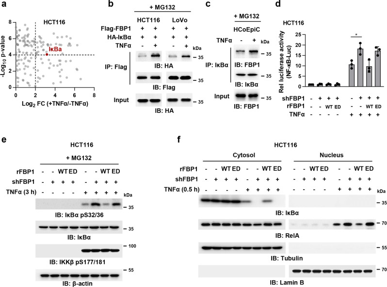
Fig. 2. FBP1 interacts with IκBα and inhibits TNFα-induced NF-κB signaling.
a HCT116 cells stably expressing Flag-FBP1 were pretreated with 50 μM MG132 for 1 h and treated with or without TNFα (10 ng/mL) for 3 h. Cells were harvested and Flag-FBP1 proteins were immunoprecipitated using anti-Flag agarose beads. FBP1-associated proteins were analyzed by mass spectrometry analyses. The data are presented as a volcano plot for two biologically independent experiments (two-tailed Student’s t-test). Proteins with P < 0.0001 and log2-transformed FC in expression of > 2.0 were regarded as the candidate proteins that showed strong interactions with FBP1. b HCT116 or LoVo cells co-transfected with Flag-FBP1 and HA-IκBα were pretreated with 50 μM MG132 for 1 h and treated with or without TNFα (10 ng/mL) for 3 h. Flag-FBP1 was immunoprecipitated. c HCoEpiC cells were pretreated with 50 μM MG132 for 1 h and treated with or without TNFα (10 ng/mL) for 3 h. IκBα was immunoprecipitated. d FBP1-depleted HCT116 cells rescued with rFBP1 WT or ED were transfected with NF-κB reporter and then treated with or without TNFα (10 ng/mL) for 3 h. The relative luciferase activities were normalized to those of the cells expressing shNT and to Renilla controls. Data represent the mean ± s.d. of three biologically independent experiments (two-tailed Student’s t-test). e FBP1-depleted HCT116 cells were rescued with rFBP1 WT or ED. Cells were pretreated with 50 μM MG132 for 1 h and treated with or without TNFα (10 ng/mL) for 3 h. Immunoblotting assay was performed. f FBP1-depleted HCT116 cells were rescued with rFBP1 WT or ED. Cells were treated with or without TNFα (10 ng/mL) for 0.5 h. Cells were harvested and cell fraction assay was performed.

