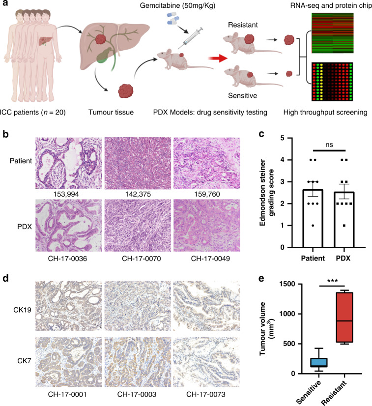
Fig. 1. Preclinical models of PDX preserved the patients’ pathological characteristics.
a The establishment of the PDX models and the screening process of research molecules in this study. b HE staining of clinical tumour tissue from patients and corresponding PDX model tumour tissue (200×). c Edmondson-Steiner grading of PDX (n = 9) and patients (n = 9) tumour tissues. ‘ns’ represents no significant difference. d Representative immunohistochemical images of CK19 and CK7 in PDX tumour tissues (200×). e Box plot of the tumour volumes at the end of treatment (day 30) obtained in PDX-sensitive tumour tissues (n = 13) and resistant tumour tissues (n = 7).

