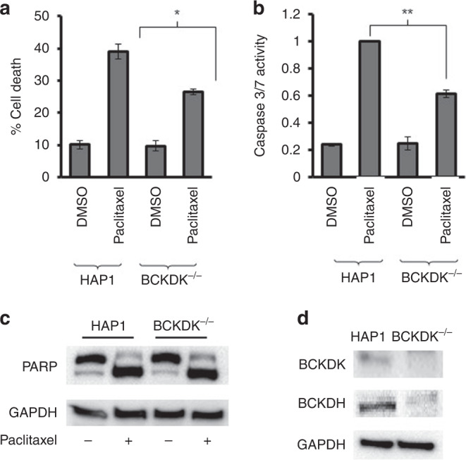
Fig. 3. The effect of paclitaxel in cells edited to lack BCKDK.

a HAP-1 and BCKDK−/− cells were treated with paclitaxel (5 nM, 24 h) and the viable cell number was determined by staining with trypan blue. The number of dead cells (mean ± S.D., n = 3) was significantly less (*P < 0.05, paired t test) in the BCKDK−/− cells compared to the parental HAP-1 cells b Caspase-3/7 activity was determined in HAP1 and BCKDK−/− cells following exposure to paclitaxel (5 nM, 24 h). The activity of caspase was normalised to relative cell number determined by SRB staining of a comparable experiment performed at the same time to control for any effect of the drug on cell number. The results are expressed as a fraction of the caspase-3/7 activity measured in the HAP1 cells (mean ± S.D., n = 3) and are significantly different where indicated (**P < 0.095, paired t test). c The indicated cells were exposed to paclitaxel (5 nM, 24 h) and PARP cleavage was determined by western blotting. GAPDH was used as a loading control. The results are representative of three experiments. d BCKDK and BCKDH were measured by immunoblotting of lysates prepared from untreated HAP-1 and BCKDK−/−. GAPDH was used as a loading control. The image is representative of three experiments.
