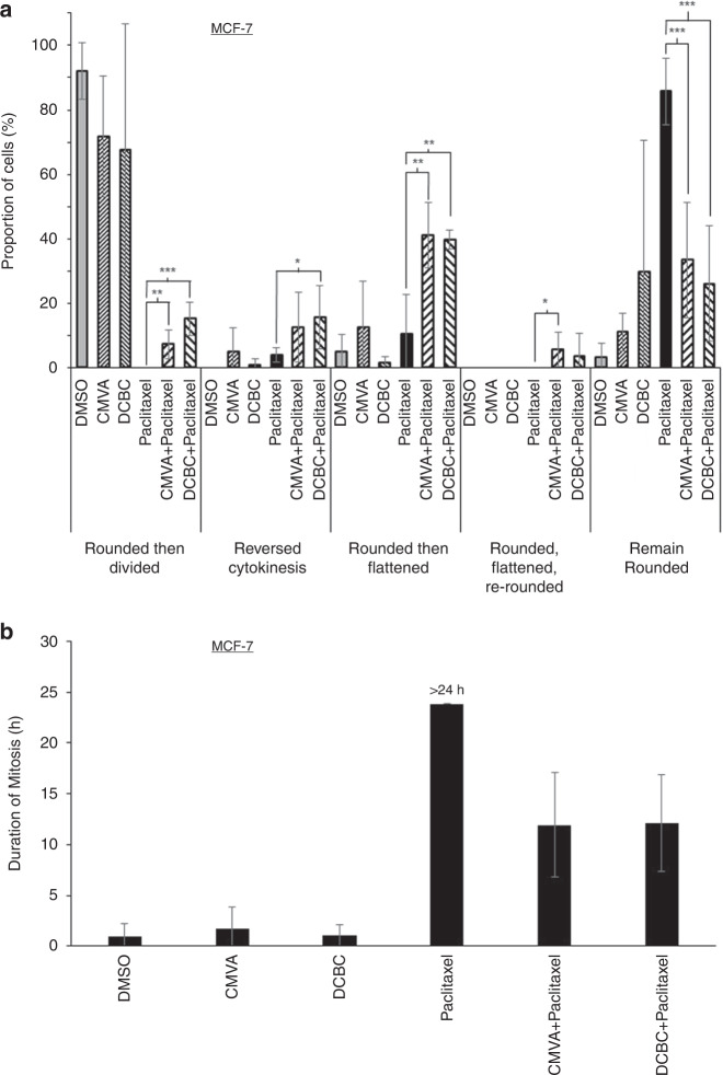
Fig. 5. Effect of BCKDK inhibitors on M-phase arrest induced by paclitaxel.
a MCF-7 cells were exposed to the indicated combinations of paclitaxel (9.5 nM), CMVA (100 µM) or DCBC (100 µM) and monitored by time-lapse microscopy. The time at which the cells rounded and their subsequent behaviour was classified (mean ± S.D., n = 3 total of 100 cells counted for each condition, taken from three separate experiments) as “reversed cytokinesis” (cells started to divide, but then aborted division), “rounded then flattened” (cells did not divide but instead reverted to a flat morphology) or “rounded, flattened then re-rounded (as the previous group, but subsequently rounded again) or “remained rounded” (the cells remained rounded until the end of the recording period). The results were significantly different where shown (ANOVA, *P < 0.05; **P < 0.01; ***P < 0.001). A similar analysis was conducted with MDA-MB-468 cells (Fig. S7). The cell fate maps for each of these experimental conditions are presented in Figs. S8–21. b The time at which cells rounded was noted and time to flatten again determined and the interval this encompassed taken to represent the time required to complete mitosis. Cells treated with paclitaxel remained rounded throughout the 24 h duration of the video and hence are labelled “>24 h”.

