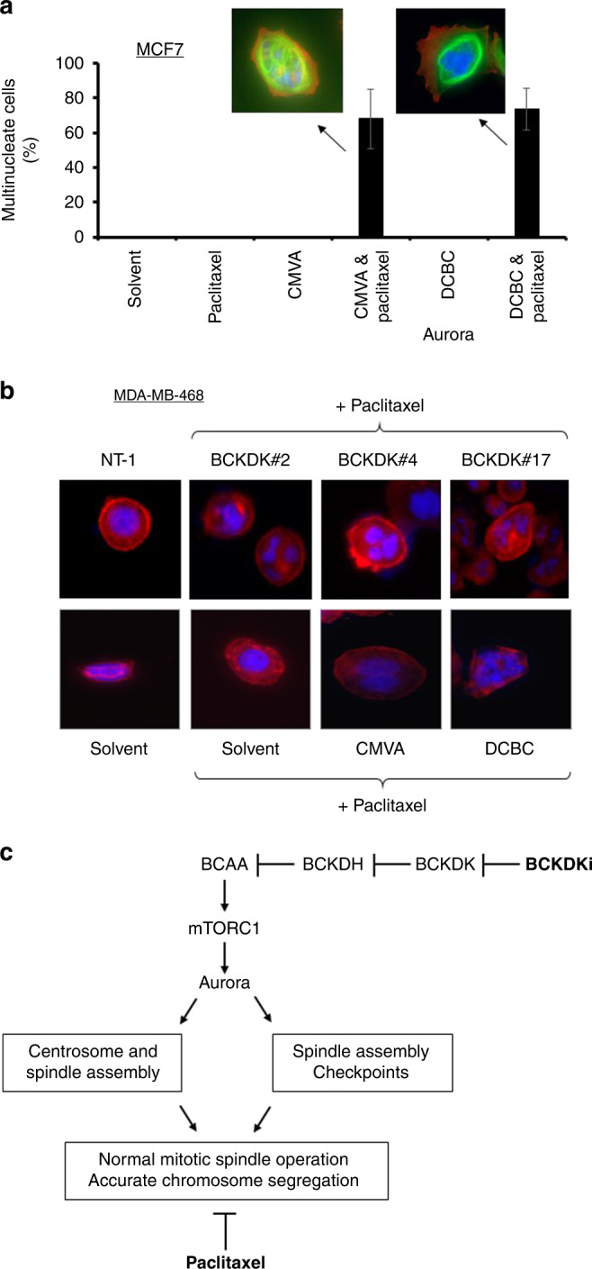
Fig. 6. BCKDKi and paclitaxel induce the formation of multinuclear cells.

a MCF-7 cells were exposed to CMVA or DCBC for 48 h, and subsequently paclitaxel and the BCKDK inhibitors for a further 24 h before fixation and staining with DAPI and anti-actin antibody. The number of multinucleate cells was determined by microscopy (mean ± S.D. n = 3, total of >100 cells counted for each condition). b MDA-MB-468 cells were exposed to the same combination of drugs or to three separate siRNA to repress the expression of BCKDK. The cells were stained with DAPI (blue) or with an antibody to F-actin (red). The number of multinucleate cells was determined by microscopy. c The proposed mechanism for augmentation of paclitaxel activity. Paclitaxel prevents the assembly of the mitotic spindle causing activation of the spindle assembly checkpoint and arrest in M-phase. Inhibition of BCKDK relieves the inhibition of BCKDH by BCKDK, thereby promoting BCAA metabolism. Activation of mTORC1 is dependent upon branched-chain amino acids so their subsequent metabolism by BCKDH leads to inactivation of mTORC1 and its substrate Aurora. This further impairs mitotic spindle assembly and/or prevent proper functioning of the spindle assembly checkpoint. Consequently, the drugs act synergistically to prevent accurate chromosomal segregation, resulting in cell death.
